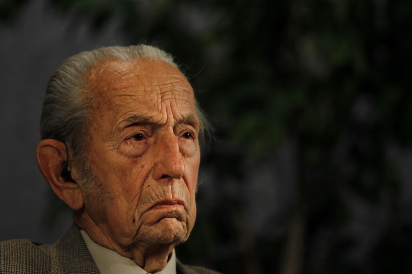
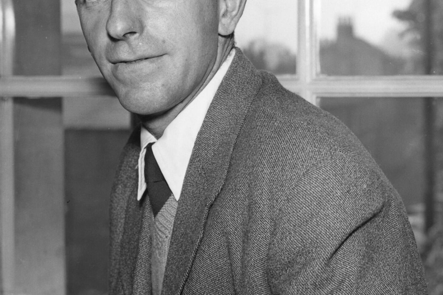
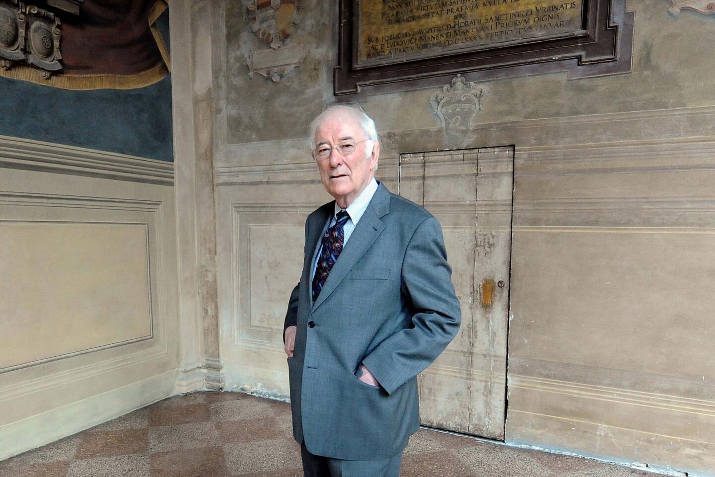
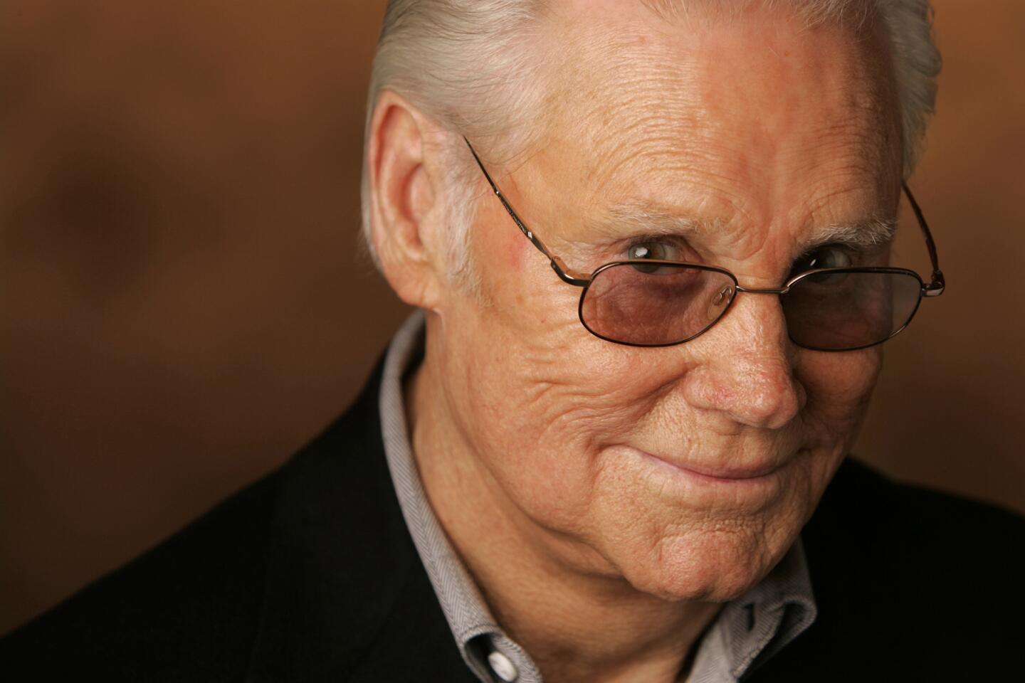
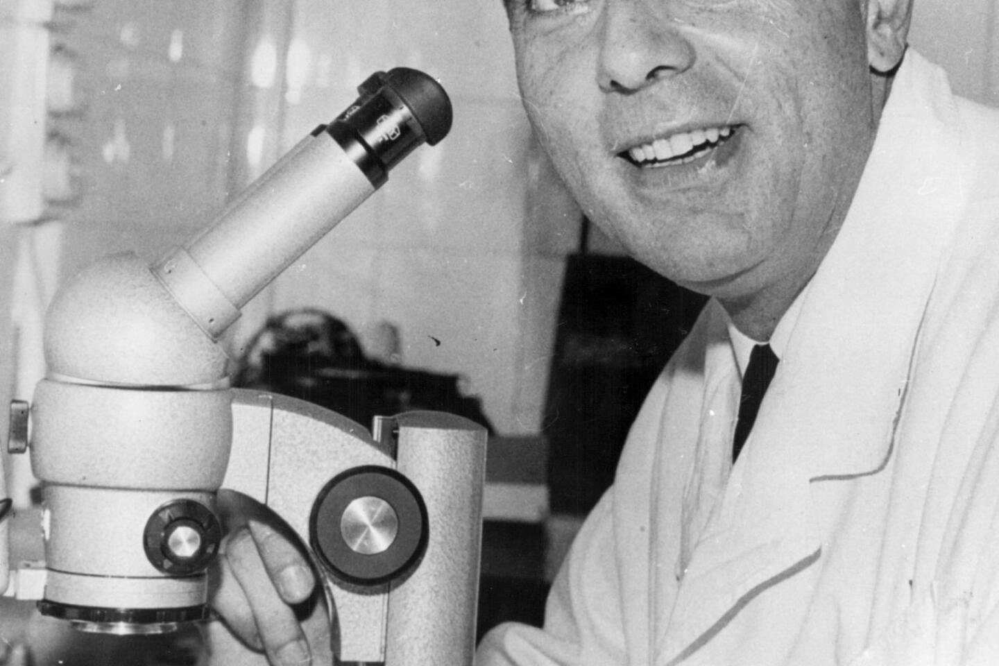
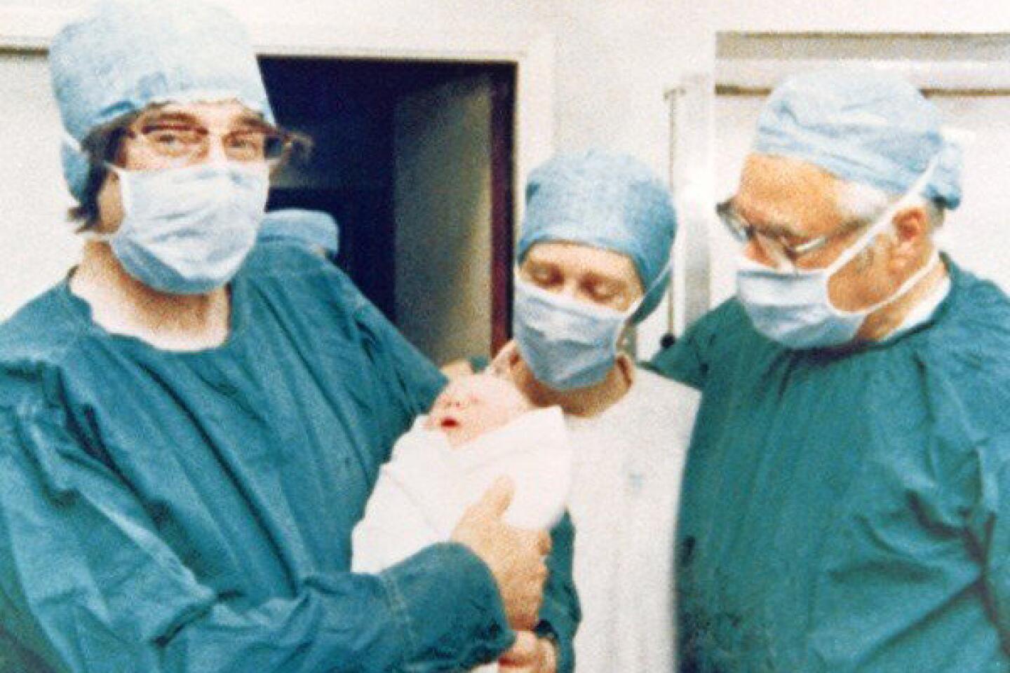
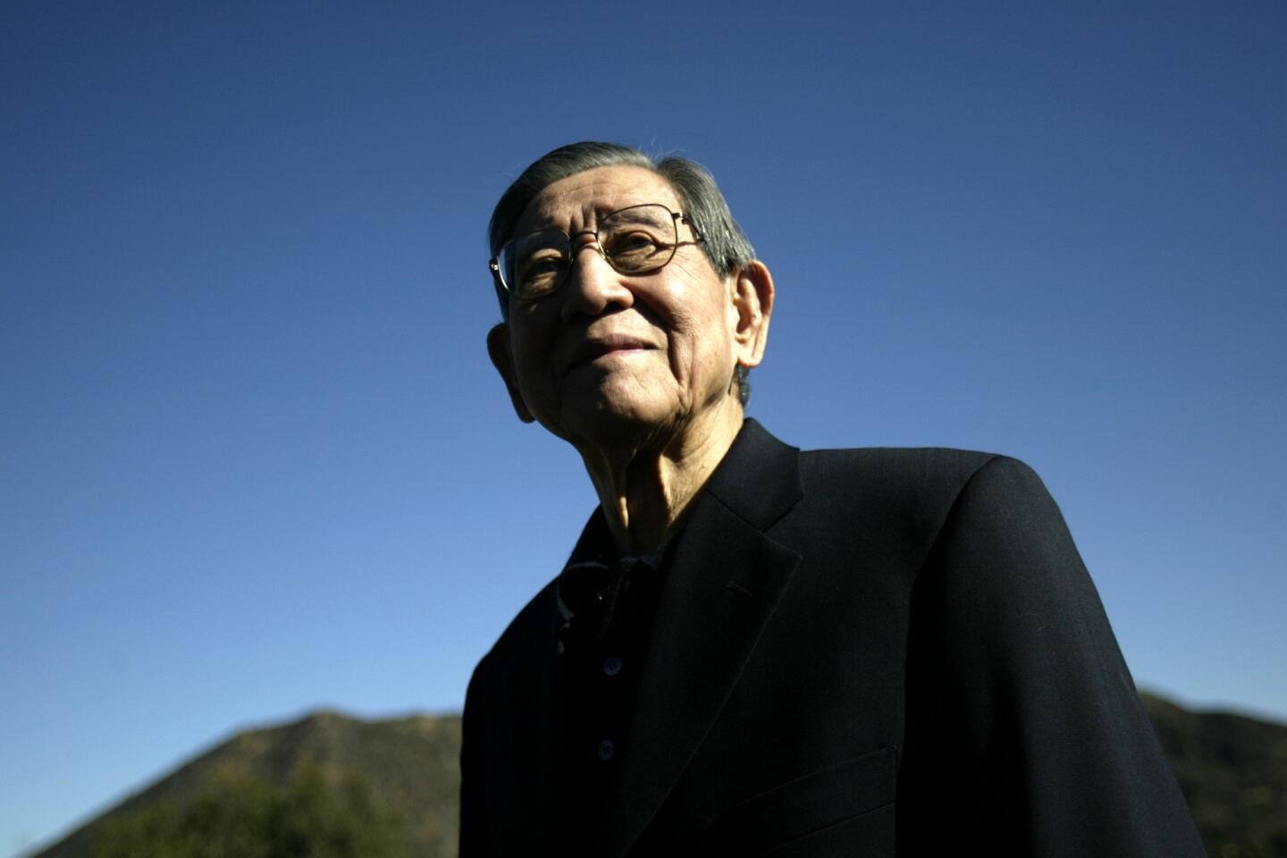
Dr. Christian de Duve dies at 95; Nobel-winning scientist


For the first half of the 20th century, the cell was a mysterious, unfathomable entity. Nutrients went in and hormones, wastes and other products came out. But what happened in between was anybody’s guess.
Light microscopes could reveal the rough details of the cell’s interior, but not with enough precision to illuminate function. Chemical studies were rudimentary at best.
Three men changed that. Albert Claude of the Rockefeller Institute — now University — adapted the electron microscope to image cells, allowing a much higher resolution. He also began the practice of what is known as differential sedimentation, in which a cell was broken up with a mortar and pestle and centrifuged, separating components by weight, and discovered the energy-producing organelles known as mitochondria.
PHOTOS: Notable deaths of 2013
George Palade of Rockefeller refined both techniques and used them to discover ribosomes, the cellular compartments where proteins are synthesized, and the endoplasmic reticulum, an extensive network in the cell that carries ribosomes and transports materials needed by the cell.
Dr. Christian de Duve, who divided his time between Rockefeller in New York City and the Catholic University of Louvain in Belgium, further refined differential sedimentation and used it to discover lysosomes, the cell’s stomach that is responsible for destroying unwanted material, and peroxisomes, which use hydrogen peroxide to destroy certain other unwanted substances.
For their efforts, the three shared the 1974 Nobel Prize in Physiology or Medicine. In awarding the prize, the Nobel committee said the men’s “accomplishments have been largely responsible for the creation of modern cell biology. What used to be a cell with components, the reality of which was often a matter of dispute and functions as a rule unknown, is now a system of great organizational sophistication with units for the production of components essential to life and units for disposal of worn out parts and for defense against foreign organisms and substances.”
De Duve, 95, died Saturday at his home in Nethen, Belgium, according to Rockefeller University. He decided to end his life with euthanasia, which is legal in Belgium. He had been suffering from cancer and atrial fibrillation, and his health problems were exacerbated by a recent fall in his home.
In a recent interview published after his death by the Belgian newspaper Le Soir, de Duve said, “It would be an exaggeration to say I’m not afraid of my death, but I’m not afraid of what comes after because I am not a believer. When I disappear, I will disappear, there’ll be nothing left.”
De Duve’s greatest discoveries were almost accidental, resulting from anomalous laboratory findings that piqued his interest and led him down new and unexpected pathways. For years, he had been interested in insulin and, in the late 1940s, was attempting to study its metabolism in liver cells.
To extract the appropriate enzymes, he ground up the cells with an electric blender and used differential sedimentation to isolate various portions of the cell. Because it was easy to monitor, he used an abundant enzyme called glucose 6-phosphatase. His initial experiments, however, indicated that destroying the cells with a blender broke them down too much, and he decided to switch to a much gentler technique.
To his surprise, with the gentler technique, the researchers found much lower levels of glucose 6-phosphatase. But when they stored portions of the isolated material in the refrigerator for a few days, they found that the activity of the enzyme increased. This suggested to de Duve that the enzymes in the cell were protected by a membrane, which slowly disintegrated in the refrigerator. This also made sense because the enzymes worked better in an acidic environment, while the interior of a cell is neutral.
“My curiosity got the better of me,” de Duve wrote in his Nobel autobiography, “and as a result, I never elucidated the mechanism of insulin.” In fact, it took researchers 30 more years to figure that out.
He concluded that the membranes that protected the enzymes formed small organelles inside the cell that he called lysosomes, or lytic bodies. Researchers have subsequently discovered at least 50 different enzymes that are contained in the lysosome.
In 1955, de Duve and Alex B. Novikoff of the Albert Einstein College of Medicine used the electron microscope to image lysosomes in partially purified cellular fractions.
The lysosome’s function is to engulf and destroy invading bacteria, toxins, unwanted cellular products and dying cells. Failure of any part of this system can have disastrous effects on the cell and the organism carrying it. De Duve showed that lysosomes play a critical role in a number of human diseases.
He identified, for example, a group of illnesses known collectively as disorders of glycogen storage, which arise when faulty enzymes in the lysosome cannot properly break down components of the cellular membranes, allowing them to accumulate in the cell. One of these, for example, called Tay-Sachs disease, is a congenital neurological disorder that typically kills its victims by the age of 5.
De Duve studied another enzyme called monoamine oxidase, which was similar to enzymes of the lysosome. It was actually located elsewhere in the cell, in the organelles that he dubbed peroxisomes because they use hydrogen peroxide to destroy foreign materials. Peroxisomes break down foreign substances, such as alcohol, and they play a role in sugar metabolism.
Christian Rene de Duve was born Oct. 2, 1917, in Thames-Ditton, just outside London. His Belgian parents had moved there to escape World War I, but the family returned to Belgium after the war. His travels to see friends in England and family in Germany, combined with his education in Flemish and French in Belgium, made him fluent in four languages.
He enrolled in the Jesuit Catholic University of Louvain, where he majored in ancient humanities. He continued in the medical school, receiving his medical degree in 1941. When World War II broke out, he enlisted in the Belgian army and became a medic. His unit was shipped to the south of France, where it was almost immediately captured by the Germans.
As the group was being marched to a prison camp, de Duve jumped on to the running board of a passing car and escaped. At that point, he concluded that he had had enough war and returned to Louvain to finish his medical studies. Deciding that he preferred research, he pursued an advanced degree in chemistry, writing his thesis on insulin.
PHOTOS: Notable deaths of 2013
After graduation, he did postdoctoral work in Sweden and St. Louis, working with four Nobel laureates. In 1947, he joined the medical school at Louvain, teaching biochemistry and performing research. In 1962, he accepted a post at Rockefeller, dividing his time between the two labs. He remained on the faculty of both until his retirement in the late 1980s.
De Duve helped found the American Society for Cell Biology and, in 1971, he created the International Institute of Cellular and Molecular Biology at Louvain. In 1997, the latter was renamed the Christian de Duve Institute of Cellular Pathology. In his later years, he focused on the evolution of life and wrote several books about the subject.
His wife of 65 years, the former Janine Herman, died in 2008. De Duve is survived by two sons, Thierry and Alain; two daughters, Anne and Francoise; two brothers, Pierre and Daniel; seven grandchildren; and two great-grandchildren.


