Doctors operate on heart of a 25-week-old fetus [video]
A wire the width of a hair. A balloon just a few millimeters wide. A needle measuring 11 centimeters exactly.
These were the tools that a team of doctors in Los Angeles used to open up a narrow aortic valve in the heart of a 25-week-old fetus still growing in its mom’s belly.
The procedure is known as a fetal aortic valvuloplasty. It was also a first for Southern California.
A few weeks before the doctors gathered in the operating room at CHA Hollywood Presbyterian Medical Center in Los Angeles, fetal cardiologist Dr. Jay Pruetz of Children’s Hospital Los Angeles diagnosed the fetus as having severe aortic stenosis. That means the baby’s aortic valve was very tight. Blood was backing up in the left ventricle of the baby’s heart, keeping it from pumping normally.
If doctors had not intervened, the left ventricle would not develop properly, and the baby would likely be born with a life-threatening condition called hypoplastic left heart syndrome (HLHS).
In the video above, you can watch part of what happened when the team of doctors did intervene.
After practicing a few times with a model of jello and a grape -- the grape standing in for the heart, the jello standing in for the surrounding body -- the doctors performed the procedure on Sept. 25. Mom had local anesthesia and was sedated. The baby was given anesthesia and a muscle relaxant so it wouldn’t switch positions at an inopportune time.
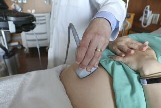
Dr. Ramen Chmait, assistant professor at Keck School of Medicine of USC and director of Los Angeles Fetal Therapy, inserted a thin metal tube (he calls it a needle) through the mom’s belly, into the uterus, through the amniotic cavity and into the fetus’s chest.
In the ultrasound video above, you can watch as he continues to maneuver the needle, ever so carefully, through the fetus’s heart, stopping when it is directed toward the narrow aortic valve.
(And for a little perspective, the heart of a fetus this young is about the size of a walnut).
With the needle in place, pediatric interventional cardiologist Frank Ing of CHLA threaded a thin wire just 14/1000s of an inch down through the needle.
The wire served as a rail for Dr. Ing to push a tiny balloon that inflates to 3.25 millimeters wide across the aortic valve. The balloon was attached to a catheter. Dr. Ing carefully inflated the balloon with a premeasured amount of liquid.
The inflated balloon stretched and even tore a small part of the aortic valve, allowing more blood to flow to the fetus’s heart.
Then out came the balloon, wire, and needle, and the procedure was over.
Both Dr. Ing and Dr. Chmait relied on ultrasound imaging by Pruetz to see what they were doing.
A few weeks after the surgery, the doctors report that both mom and fetus are doing fine.
“It’s only been a week or two, but even initially after the procedure, we could see increased blood flow across the valve, and the heart was squeezing a bit better than before,” Dr. Pruetz said.
“There is no question in my mind that without this procedure the baby would have had HLHS,” Dr. Chmait said. “Now the baby has a chance to have the left ventricle recover with some good function.”
The procedure was orchestrated by a consortium of fetal therapists with the CHLA-USC Institute for Maternal-Fetal Health at Hollywood Presbyterian.


