Poisoned pets, preemie foals, dogs with spina bifida: UC Davis’ top-ranked vets see them all
- Share via
A Saudi royal cat was flown in for a kidney transplant. Bulldogs with spina bifida come for stem cell therapy.
Lame horses arrive to get diagnosed using a scanner found in no other animal hospital.
A dog who became a hero in the Philippines when she threw herself between a speeding motorcycle and two children was brought here because her upper jaw and snout had been torn off in the crash.
You never know what will come through the doors of the UC Davis Veterinary Medical Teaching Hospital, a showcase of pioneering therapies, advanced technologies and elite researchers. The top-ranked veterinary program in the world offers 34 specialties including cardiology, oncology and neurology.
More than 50,000 animals a year — as small as hamsters, as big as buffalo — are cared for by its 120 faculty veterinarians and nearly 600 interns, residents, fellows, technicians and students.
Its specialists assist endangered gorillas in Africa, penguins in Brazil — even residents, including tarantulas and giraffes, of the nearby Sacramento Zoo.
The hospital recently launched a $508-million project to double its size in 10 years, which would make it one of the world’s largest veterinary hospitals.
“It’s a real gift to have a world-famous vet hospital an hour away,” said Mary Shallenberger of Clements, who brought in her 4-year-old quarter horse, Oscar, for a spinal tap after she noticed weakness in his hindquarters. He has since made a complete recovery.
Here’s a look at some of the animals in need and the doctors who cared for them.
Stem cells for Spanky and Darla
Spanky and Darla bounded into the exam room, all floppy-eared and wrinkly-faced. The English bulldogs seemed perfectly normal — except for their diapers, held up by animal-print suspenders. Brother and sister, they were born with spina bifida, and their improperly formed spinal columns made them incontinent and not so steady on their feet. The dogs’ owner planned to euthanize them. Val Vallejo, a Los Angeles County sheriff’s deputy who runs the nonprofit Southern California Bulldog Rescue, brought them to UC Davis instead.
At UC Davis, Darla and Spanky became medical pioneers when the medical and veterinary schools teamed up in February to provide them with the first-ever spina bifida treatment for dogs that combined surgery and stem cells.
A team led by Dr. Beverly K. Sturges, a veterinary neurosurgery professor, repositioned misplaced tissue around the dogs’ spinal columns. Then specialized placental stem cells developed by Drs. Aijun Wang and Dori Borjesson, professors and stem cell researchers, were applied to cover the gaps in the spinal columns and promote regeneration.
The dogs still need their diapers. It’s unclear whether that issue will ever be cleared up.
But, said Vallejo, who drove all night for the recent check-up, “Their gait looks better.”
And so do their prospects. Darla and Spanky, Vallejo reported, are happy and living with a permanent family in New Mexico.
Advanced scans for Scooby
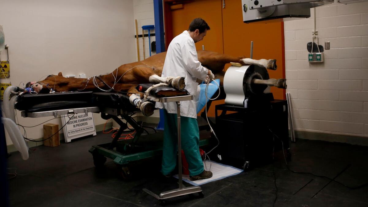
Scooby is 15 years old. He’s a chestnut-colored German warmblood horse. His specialty is dressage. He’s an expert in show riding. But when he began limping in March, no one at first knew where the trouble was.
In April, UC Davis veterinarians hoisted all 1,300 pounds of him onto an examining table and placed tubes for oxygen and anesthesia gas in his mouth. His right hind leg poked though a wide circular tube in a positron emission tomography, or PET, scanner. UC Davis was the first veterinary medical hospital anywhere to use such a machine on horses.
By injecting the leg with a weak dose of a radioactive substance that attaches to areas of abnormal bone, the veterinarians were able to see detailed images of Scooby’s injury.
“This can detect lesions missed by MRI and CT scans, which could prevent catastrophic breakdowns in racehorses,” said Dr. Mathieu Spriet, an associate professor of surgical and radiological sciences. And the low-level radiation doesn’t hurt the horse.
Scooby’s scans found a small crack that led to inflammation nearby. After months of medication and rest, Spriet said, he is back to being ridden at a walk and trot pace and appears to be on the right track.
Jawbone surgery for Gigi
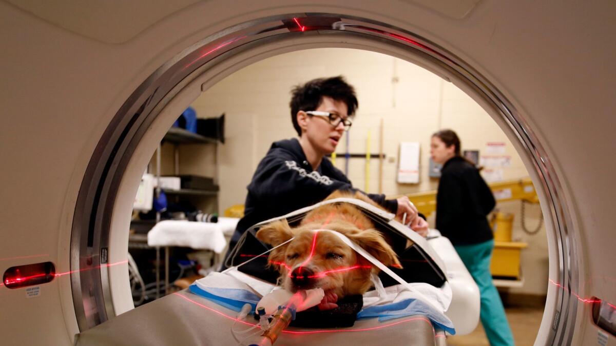
Gigi, an 8-year-old mixed-breed dog, was strapped down on her belly, ready for a CT scan. A local vet had removed a tumor in her jaw, but it had grown back into the bone. Enter Drs. Frank Verstraete and Boaz Arzi, two UC Davis veterinary oral surgeons famous for developing, with the school’s biomedical engineering department, the first procedure to regrow jawbones in dogs.
The surgeons can reconstruct removed sections of a jawbone using a titanium plate and screws, and then pack them with a material soaked in a protein that adheres to the original bone and stimulates regrowth. To shorten the time a dog has to stay under anesthesia, they make 3-D models of each jawbone and prefit the plate and protein material before surgery. They have successfully treated more than 30 dogs, including Frankie, a spaniel who was found in a Marin County forest by campers after he had been shot in the face and left to die.
In Gigi’s case, the surgeons peering at scans concluded that the tumor was small enough to avoid a major amputation and jawbone reconstruction for now. She seemed to be doing fine after she underwent a more limited surgery to remove the tumor and some teeth and bone.
Taking a stand for Danika
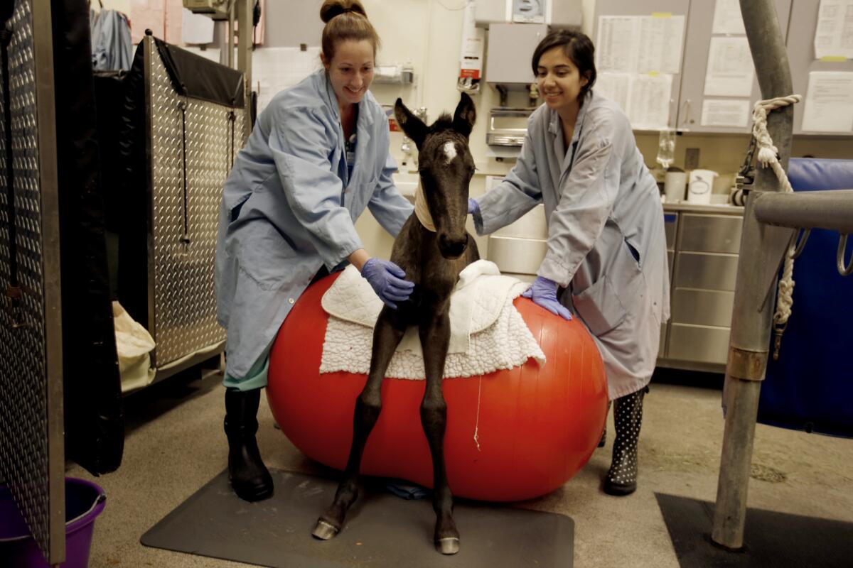
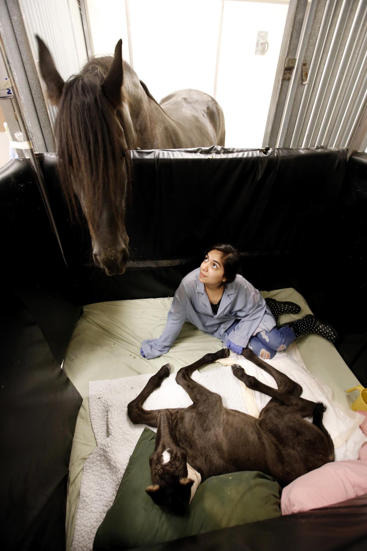
Danika arrived four weeks prematurely, a Friesian foal the color of coffee, with a milky-white star on her forehead. She was born so early that her bones had not fully formed. So at UC Davis, LuzMaria Soto, studying animal science, sat next to the horse to keep her from trying to stand up on her own. If she did, she could damage her bones and wind up permanently lame.
Danika was in good hands at the hospital’s Neonatal Intensive Care Unit. The team there is used to treating serious problems, including ruptured bladders and sepsis, said Dr. Gary Magdesian, a professor of equine critical care.
Soto and other volunteers with the UC Davis Foal Team stay with preemies around the clock.
The unit features adjoining stalls, so Danika’s mama, Rixt, was able to stay close, peeking over the divider with a watchful eye.
Every two hours, the volunteers let Danika up for assisted standing. Every four hours, they helped her bear weight by straddling a bouncy exercise ball. The foal also was given enzymes to treat a gut problem. After five weeks in the NICU, Danika was declared good to go.
Better blood for Crystal
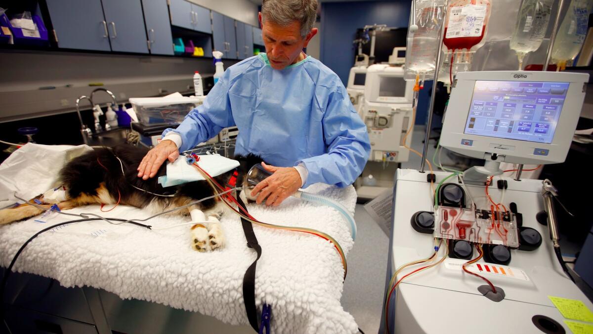
Crystal, a 4-year-old Australian shepherd, had become lethargic. Her gums were pale. Dr. Larry Cowgill explained that the dog’s immune system “got confused” and was destroying red blood cells faster than it was producing them.
So Cowgill prepared Crystal for one of his pioneering blood purification therapies. Called therapeutic plasma exchange, the treatment removes plasma tainted with damaging antibodies, toxins or abnormal proteins and replaces it with healthy donor plasma. The plasma is separated from the blood by a centrifuge. Cowgill’s team hooked Crystal up to the machine’s two blood lines, then hit the start button. By the end of the 30-minute treatment, the veterinarian said, he expected Crystal’s red blood cell count to go up from 16% to 30%, closer to the normal range of 40% to 45%.
The Hemodialysis and Blood Purification Unit that Cowgill heads is the largest in the country, performing 340 treatments last year. Most of the patients are dogs who may have gobbled up something they shouldn’t — say antifreeze or ibuprofen. “It’s always a Lab retriever,” Cowgill joked — though cats sometimes eat toxic lilies.
The blood treatment helped turn Crystal around within a week.
“It’s magic for these animals,” Cowgill said.
Twitter: @teresawatanabe
