Sticky Issue of Sea Urchin May Offer a Clue on Cancer : ‘Glue’ Binding Cells Together Yields Insights Into Life’s Basic Processes
- Share via
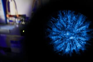
After years of painstaking experiments on sea urchins, a biologist at California State University, Northridge and a team of students have isolated a molecular “glue” that makes the cells of sea-urchin embryos stick together.
Studying the stickiness of sea-urchin cells might seem an esoteric pursuit. But the biologist, Steven Oppenheimer, said the work should lead to insights into pressing questions such as how some birth defects occur and why cancer cells sometimes break loose from a tumor and malignantly spread in the human body.
Generous grants last year from both the National Science Foundation and National Institutes of Health attest to the importance of the sea-urchin studies, said Oppenheimer, who has 20 students working on the project.
A Fundamental Question
“In fact, stickiness is one of the tremendously fundamental questions in all the life sciences,” Oppenheimer said, as he paced the cluttered confines of his laboratory in the basement of the campus science building.
If the individual cells in a human body or that of any other organism did not stick together, he said, all life forms would have the consistency of soup. As an organism grows from embryo to adult, specialized tissues appear--groups of liver cells, skin cells, heart muscle cells and the like--each cell somehow “knowing” what its neighbors should be.
Despite the pervasiveness of this seemingly simple phenomenon, surprisingly little is known about what makes cells adhere to each other, he said.
When something about this stickiness goes awry during embryonic development, a birth defect may be the result. And in an adult organism, the tendency of cells to stick together or slip apart plays a crucial role in cancer, said Oppenheimer, who, when he is not in the laboratory, is raising funds for CSUN’s Center for Cancer and Developmental Biology, which he directs.
“In cancer, the stickiness is messed up,” he said. When a tumor becomes malignant, something about the surface of individual cancer cells changes, allowing them to become unstuck from their neighbors.
When Cells Become ‘Unglued’
If a cancerous tumor just grew in one place, it could easily be spotted and surgically removed. But when a cancer becomes malignant, its cells become “unglued” and spread around the body, invading and damaging various tissues. “The altered stickiness of cancer cells is probably one of the most important reasons why cancer is dangerous,” he said.
Unfortunately, he said, “It’s difficult to study the biology of cancer by looking directly at cancer. You’re dealing with very, very complex cells, in environments that are difficult to reproduce experimentally.”
That is where sea urchins come in. Various alternatives that provide a laboratory model of cell-to-cell stickiness have been looked at by researchers--including cultures of cells from the retinas and muscles of chickens--but none present such an ideal system for studying cells’ interactions, Oppenheimer said.
Simple Embryos
The embryos of sea urchins, unlike those of say, a mouse, are simple systems, he said. They also can be raised easily in their natural environment--seawater. In addition, they can be produced in abundance.
“One of the biggest problems with this kind of work is getting enough experimental material,” Oppenheimer said. Embryos of mammals or animals high on the evolutionary ladder can only be gathered in small numbers, but “for biochemical analysis, you need literally quarts of these cells,” he said.
Rolling up the sleeve of his laboratory coat, Oppenheimer pulled two sea urchins from an algae-green aquarium. This is where the experiments begin, as the adult urchins are stimulated with a chemical to release streams of millions of eggs and sperm.
Soon after the eggs are fertilized, they begin to divide, subdivide and grow into embryos. “We can easily raise as many as we need,” Oppenheimer said.
Studied at Early Stage
The embryos are studied at the blastula, an early stage of development where they are transformed from a loose collection of several hundred cells into a swimming organism with a distinct shape.
Oppenheimer took a flat glass dish full of sea-urchin embryos out of a refrigerator. Bathed in seawater in the dish, they were barely visible, resembling a sprinkling of the finest beach sand. But under a low-power microscope, each grain could be seen as a tumbling, translucent organism with a body shaped like a pitted olive.
In 1982, Oppenheimer discovered that when sea-urchin blastulas were placed in seawater from which two elements--calcium and magnesium--had been removed, the embryo fell apart into hundreds of individual cells and gave off “sticky stuff”--particles that seem to glue the cells together.
Cells Regroup
Remarkably, when a loose soup of these cells was returned to normal seawater and mixed once again with the “sticky” particles, the cells came together and built themselves back into nearly normal embryos--in a process as unlikely as a pile of bricks building itself into a wall. Although the rebuilt embryos would not mature into sea urchins, they swam and appeared nearly normal, he said.
It is those sticky particles--given the name “adherons” by Oppenheimer--that are at the heart of the research project. The work has attracted grants from NIH and other sources totaling $300,000 over the last five years--a considerable amount, especially for a university that is not a major cancer research center. Oppenheimer’s group has used the money to begin to isolate proteins on the adherons that seem to be the adhesive.
Purification and Analysis
Last week in the laboratory, two of Oppenheimer’s students, both pursuing master’s degrees and applying to medical school, were purifying and analyzing the adherons. Through a series of chemical steps, the particles are broken into their constituents. The soup of ingredients is then placed on a plate of gelatin in an electric field, which tugs on molecules of various sizes, pulling them various distances through the gelatin. This helps to identify molecules by their weight and electrical charge.
Many questions remain about the chemical structure of the molecules and how they interact with the surface of cells, Oppenheimer said. How is it that adherons from one species, the brown sea urchin, do not work with embryos of another species, the purple sea urchin? Do embryos of other organisms have this cellular glue? Is there a similar process occurring in tissues in adult organisms?
Even after 15 years of work on sea urchins, Oppenheimer readily admits the possibility that there may be no clue to the workings of cancer hidden in this mass of questions. But he is confident that he is on the right track. “Many of the major breakthroughs in science have been made as a result of little experiments . . . that no one would ever have thought were significant,” he said.
SEA URCHIN LINK IN CANCER RESEARCH ADULT SEA URCHIN
Sea urchins are the focus of research at CSUN that may explain how cancer cells can break loose from a tumor and spread around the human body. Scientists there have found tiny protein particles that act like glue, holding the many cells in a sea urchin embryo together.
BLASTULA
Like other organisms, early in life, sea urchins pass through a stage of development called a blastula. Each cup-shaped blastula contains just a few hundred cells. In seawater, the embryos swim and continue to grow, eventually reaching the spiny adult form. CELLS BECOME “UNGLUED”
But when a blastula is placed in seawater that has had certain elements removed, it becomes “unglued,” breaking into individual cells and “sticky” particles, which the scientists have called adherons. This may mimic the process by which a tumor becomes malignant. BLASTULA BEING REBUILT
When placed back in normal seawater and mixed with the adherons, the cells somehow reconstruct themselves into a nearly normal sea urchin embryo, which is capable of swimming. The research team is trying now to identify the chemical structure of the adherons.


