PERSONAL HEALTH : New Focus on Key Test for Cancer : Radiology: Mammograms can provide vital early warnings of breast cancer, but some investigation of facilities is necessary to ensure that the tests are properly conducted and results are reliable.
- Share via
Recent concern about the accuracy of Pap tests not only has underscored the need for greater care with that type of cancer detection. It also has given new relevance to a quiet effort to improve the accuracy of one of the few other mass-screening tools for cancer: mammography.
Unlike the Pap test, the mammogram offers women choices and a competitive market in which to make them.
But until recently there has been no easy way for a woman to know that the facility she chooses will detect problems early enough to prevent her from being among the 43,000 women who die every year from breast cancer.
In fact, radiologists concede, faulty procedures and inadequate quality control in some cases have resulted in poor-quality mammograms that miss small cancers.
No one can say how many women may have been given a false sense of assurance when in fact they had cancer, but “we’re certain” it has happened, said Dr. Lawrence W. Bassett, director of the Iris Cantor Center for Breast Imaging at UCLA.
How can a woman know if her mammography facility is a good one? “The answer,” he said “is that until recently we really had no mechanism. Women were told . . . that if they asked certain questions they could find out. But that puts an awful lot of burden on women. You shouldn’t have to do an investigation on your own every time you use a service,” he said.
Bassett is helping to lead a national push by radiologists, the government and the American Cancer Society to remedy the problem by setting standards for quality mammography.
Unlike previous concerns over excessive radiation doses--concerns focusing on old or poorly adjusted equipment--the latest effort aims at improving the more subtle procedural aspects of X-raying a woman’s breasts.
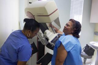
Getting a quality image while keeping the radiation dose down requires: well-adjusted equipment dedicated solely to mammography; extended processing of the film separate from other X-rays; well-supervised technicians; and proper positioning of a woman’s breast against the X-ray film, Bassett said.
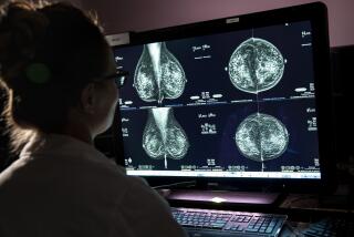
“If the image that comes out is good, it’s not only because the equipment’s good, but because the technologist positioned the patient properly, the compression (of the breast) was adequate, and the radiologist knew how to read it,” he said.
The keystone of the new push is a voluntary certification program by the American College of Radiology. Since its inception two years ago, the program has certified about 2,000 facilities as meeting its standards for taking and interpreting mammograms; 52 of these are in California, with another 59 in the state in the process of certification, said Marie Zinninger, who directs the program.
“This is meant to be an educational effort, to raise a level of awareness about what needs to be done to have quality mammography,” Zinninger emphasized.
As a result, facilities that don’t pass usually adjust procedures or equipment and reapply successfully, she said.
The government also is getting into the act. In California, the radiologic health branch of the Department of Health Services has drafted rules to require mammography facilities to regularly test their work to assure quality control, said Edgar Bailey, branch chief. It also will adopt rules of good practice for mammography.
Nationally, the Medicare catastrophic care bill passed by Congress last year would have extended mammography benefits for the first time under a federal program. It also set up strict standards for the procedure similar to those advanced by the American College of Radiology.
Since Medicare regulations often become the norm throughout the health care industry, mammography advocates were elated at what they saw as a first step toward making the test a routine part of health care covered by private medical insurance.
But mammography coverage may be excluded in the current effort to repeal catastrophic health care. The House voted Oct. 4 to repeal the entire catastrophic care bill. Mammography coverage was one of a few provisions that the Senate voted Oct. 6 to retain, while repealing most of the bill, said Ray Gill, a spokesman for the Health Care Financing Administration. The difference is being resolved in a conference committee.
For a woman seeking a mammogram, the national focus on quality will, in the future, make it easier to have confidence that her test will be properly conducted.
But for now, there is still an element of investigation necessary.
The American College of Radiology’s program, so far, has certified only about a third of 6,400 mammography facilities known to be in existence in 1988. (Zinninger estimates the number may have risen now to as high as 8,000.)
American Cancer Society offices have lists of accredited facilities in their areas. State-by-state lists of accredited facilities also are available from the federal Cancer Information Service, (800) 4-CANCER (800-422-6237).
If there are no accredited facilities nearby, women still must do some investigating to ensure the quality of their mammogram. But by making some relatively simple inquiries, they can greatly increase their chances of undergoing a test that results in good-quality images with minimal radiation exposure, Bassett said.
Radiologists note that the lowest dosage isn’t always the best. That’s because a dose that is too low means an inadequate X-ray, radiologists say.
According to the federal Center for Devices and Radiological Health, the average radiation dose from a single mammogram has decreased from unacceptable levels as high as 6 1/2 rems in 1974 to averages of 1/4 to 1/2 of a rem in 1985. (A rem is a measure of the biological effect of radiation; a chest X-ray is about 0.030 of a rem--more commonly expressed as 30 millirems, or thousandths of a rem. By comparison, cosmic rays and other environmental sources of radiation deliver 100 to 200 millirems to each American annually.)
In a 1988 national survey by the Food and Drug Administration’s Center for Devices and Radiological Health, however, new imaging techniques that can spot smaller cancers had increased the average mammogram dosage very slightly. That is because these techniques require higher energy to produce an image.
The same study found that the quality of American mammography facilities--measured by having them X-ray “phantoms” embedded in opaque material--had improved as much as 22% between 1985 and 1988.
Still, the American Cancer Society advises that a mammography test involving two views of the breast should deliver no more than 1 rem of radiation, notes Dr. Judy Dean, a Santa Monica radiologist.
Dean said doses that are 20% of that can easily be achieved. Women needn’t worry about radiation dose if the facility they or their doctor selects meets the American College of Radiology standards, she suggested. (Her own mammography center is undergoing certification.)
More important is getting women to come in for mammograms at all, she said. Data from the 1987 National Health Interview Survey showed that only 37% of women 40 and older have ever had a mammogram.
A coalition of 11 groups, including the National Cancer Institute and the American Cancer Society, united earlier this year to endorse mammograms every one to two years for all women ages 40 to 50, and annually for all women older than 50.
There appears to be agreement on that as a minimum standard, but some doctors--including Bassett and the 65,000-member American College of Physicians--contend that mammograms should be done annually on all woman older than 40.
This, they say, is necessary because cancers are harder to detect, yet more likely to be of the fast-growing type in a premenopausal woman’s dense breast tissue. As women age, this glandular tissue is replaced by fatty tissue that is easier to image.
Dean said she thinks she knows why women don’t get mammograms: “In some ways, it’s the same thing that keeps them from going to the dentist--unpleasant, easy to put off. But if you go to the dentist what’s the worst thing you can get? Maybe a root canal. If you go to get a mammogram what’s the worst thing you can hear? ‘You have breast cancer. You might die.’ It’s a big anxiety.
“I get patients in here so nervous, sometimes they arrive and they’ve been drinking. They’re half-drunk because they couldn’t face up to it otherwise,” Dean said.
What does she tell women who are fearful? “Getting a mammogram 99% of the time means getting good news. And if you’re coming regularly, in my opinion you’re never going to get the worst news. You’re going to at most find out that you have something early that needs attention but is probably curable.”
HOW TO CHOOSE A MAMMOGRAPHY FACILITY
The American College of Radiology’s program has certified only about a third of 6,400 mammography facilities known to be in existence in 1988. To get the list, call the Federal Cancer Information Service at (800) 4-CANCER.
If there are no accredited facilities nearby, the American College of Radiology advises women to choose one that can answer “yes” to all the following questions, because such a facility is likely to produce good images with minimal radiation exposure.
* Is the radiologist certified by the American Board of Radiology?
* Are technologists certified by either the American Registry of Radiological Technologists or the state licensing board?
* Is the X-ray equipment dedicated solely to mammography or specifically designed for it?
* Is the equipment calibrated regularly by a certified radiological physicist? (Once a year is considered minimum.)
* Have the radiologist and the technologist taken special courses or had additional training in mammography?
* Does the radiologist do mammography as part of his or her regular practice? Source: American College of Radiology


