HEATH HORIZONS : MEDICINE : Mammography--Vital but Not Foolproof : IT IS THE ONLY TECHNOLOGY CAPABLE OF FINDING BREAST CANCER IN ITS EARLY STAGES, WHEN THE CURE RATE IS 91%. BUT THERE ARE INHERENT LIMITS, ESPECIALLY FOR WOMEN UNDER 50.
- Share via
Mary Stupp had her first mammogram in 1987 when she was 57. It read: “normal.” A few months later she felt a small lump in her breast, but waited to have it checked during her next annual mammogram. Once more she was reassured. It read: “no malignancy.”
But when her breast skin dimpled, the professor of philosophy at Mountain View College in Dallas decided to see her internist instead of going back to the mobile facility that had taken her previous mammograms. Her internist read reports from the two exams but did not view the mammograms himself. He gave her a clinical exam during which, Stupp says, he felt the lump. He assured her that there was nothing to worry about.
When Stupp went back to her physician for her annual mammogram a few months later, a suspicious-looking lump appeared on the film. Two days later, Stupp’s breast was removed. The tumor was the size of a lemon.
When Stupp finally managed to get her original mammograms from the mobile facility (operators first said the X-rays were lost, she said, and later sent them), the two X-rays showed clear evidence of suspicious-looking abnormalities. Now ill with inoperable cancer that has spread throughout her body, Stupp is dying. Her family sued the radiologists who read her first two mammograms.
Ruth Green (a pseudonym) of Sacramento had a more positive experience. Green was 47 when she went to her family doctor for a general check up and a mammogram. Something suspicious appeared in her X-ray, and a second mammogram was taken that clearly revealed a cancerous looking lump in her left breast.
After a biopsy confirmed that the lump was malignant, Green’s breast was removed along with a tumor the size of a golf ball. Doctors believe they caught the cancer early enough and expect her to live a normal life span.
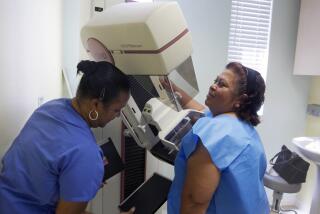
The polar experiences of these two women demonstrate some of the problems and drawbacks of the science and application of mammography. Mammography is the only technology capable of finding breast cancer in its early stages--two years before a woman can feel it with her hand and before it has spread beyond the breast into the lymph nodes. At this stage, a cancerous growth is about the size of a garbanzo bean and has been growing about five years.
The advantage and importance of early mammographic detection cannot be exaggerated. There is a 91% cure rate when cancer is found at this point. Cure rate in this context means that doctors expect a woman to die of causes other than breast cancer.
But mammography, an X-ray of breast tissue that shows potentially cancerous shadows, is fallible and has inherent limits. About 10% of breast cancers cannot be detected in mammograms. In women under 50, whose breast tissue is dense and lacks fat, mammograms fail to spot about half of breast cancers, according to the December issue of Consumer Reports on Health. In women age 50 and older whose breast tissue is very fatty and where mammograms are most successful, mammograms may still miss one-tenth of cancers.
Then there are tragic incidents unrepresented by numbers, like that of Stupp, where a mammogram that shows some abnormality in breast tissue is not followed up with a biopsy or further tests.
Mammograms alone are not reliable and accurate enough to diagnose cancer. Even when mammograms detect an abnormality, 90% of abnormalities that cannot be cleared as benign findings from the X-ray turn out to be benign after a biopsy is performed.
In the general screening population (women going for regular screenings) false-positive readings (abnormalities followed by biopsies that are benign), range between 60% and 70%, says Dr. Lawrence Bassett, director of UCLA’s Iris Cantor Center for Breast Imaging. Bassett says false-negative readings occur about 10% of the time.
Despite this margin of error, mammograms are the most technologically advanced method used for screening breast cancer. Magnetic resonance imaging, an expensive and lengthy screening process used for brain, spine and muscular skeletal X-rays, is still being explored as a detection device for breast cancer at the UCLA center and at Baylor University in Texas. MRI uses magnetic fields and radio waves to get an image of the body.
This year, breast cancer will take the lives of 44,500 women and occur in 175,000 more women. It is the second-leading cause of death of American women after lung cancer. It will invade one in nine women during their lifetime--up from one in 11 in 1980. Experts say the value of early detection is that it could save the lives of 30% of women who might otherwise die of the disease.
Indeed, against the backdrop of such daunting statistics, mammography’s advances in the field of cancer treatment and its potential for further diminishing breast cancer deaths are encouraging. But some radiologists say doctors and patients alike are susceptible to a false sense of security when mammograms come back with an all clear. In the case of Stupp, her internist never examined the original mammograms for abnormalities. He relied solely upon the inaccurate reports that read: “normal” and “no abnormality.”
“Women come to find out that their mammogram is negative and a certain amount of assurance should go along with that, but not to the extent of excluding the (breast self-exam and clinical) tests,” said Bassett, a leading proponent of mammographic quality control and chairman of the American College of Radiologists Commission on Breast Imaging.
The American Cancer Society and the National Cancer Institute recommend that women undergo self-exams, exams by doctors and mammograms done at regular intervals throughout their lives (see accompanying story). They should never rely on one alone, or take mammography for more than it is--a fallible detection device.
Clearly, mammography is an extremely sensitive radiologic process that, if carried out with inaccurate equipment and poorly trained technicians, can be inaccurate. All the more reason for stricter regulation and higher standards, experts say.
Only nine states, including California, have comprehensive mammography control, with about 40% of the estimated 10,000 mammography machines in the nation meeting voluntary accreditation standards set by the American College of Radiology. In the last couple of years, this general lack of regulation has spawned a call by breast cancer activists, the American College of Radiologists and the U.S. Centers for Disease Control for federal and state legislation regulating mammography facilities.
What exists is an amalgam of regulation by state, federal and private agencies. California legislation requiring state inspection of mammography machines yearly became law in January. (Prior law required state inspection every three years.) And a year-old federal law strictly regulates mammography facilities screening Medicare recipients, so that poor women are not priced out of high-quality breast care.
The American College of Radiology has a 4-year-old voluntary accreditation program that, among other things, requires that machines be tested for accuracy by taking an X-ray of a model human breast that has tumors, breast disease and micro-calcifications (tiny groupings of breast calcifications that sometimes signify cancer).
But what proponents like Bassett have pinned their hopes on is the proposed federal Breast Cancer Screening Safety Act, which was introduced by Sen. Brock Adams (D-Wash.) in October 1990. The bill would require that facilities meet equipment and personnel quality standards with yearly inspections conducted and recertification every two years. Federal sanctions would be imposed on violators. Congress will examine the bill again in March. The bill was referred to the Senate Labor and Human Resources Committee last year to be rewritten and fine-tuned as suggested by breast cancer organizations.
Medical professionals, regulators and cancer activists alike agree that the issue of mammographic quality control is a serious one. During a 10-month period last year, 14 mammography machines in Los Angeles County out of about 100 inspected were closed down by the county Radiation Management Department of Health Services for violating the California Radiation Control Regulations. (There are about 325 machines countywide). The machines closed down were screening an estimated 4,300 women a year.
The majority of violations had to do with poor image quality, which increases the potential for an incorrect reading of breast tissue. Unlicensed technicians and the use of mammography machines not specifically designed for breast X-rays were also cited.
“There really is a broad range,” said Kathleen Kaufman, director of Radiation Management for the county Department of Health Services. “We have actually requested facilities stop taking mammograms immediately, where the quality was poor. Then there are facilities where the quality is so high that we as inspectors don’t hesitate to go get our own mammograms done there.”
Kaufman believes county closure of 14 facilities out 100 reflects the rate of mammographic quality nationwide.
Mammogram quality is not the only problem in detection, however. The prospect of breast cancer is still so frightening that many women hesitate to get a mammogram. As Pasadena technician Brenda Miller puts it: “There is a lot of fear and anxiety over the exam and, of course, there is a lot of anxiety over the possibility of the outcome. We get women in their 70s, 80s and even 90s who have never had mammograms, mainly because their doctors never told them to get one. I do think women need to ask more questions. They leave it all up to their doctors. They don’t do it till their doctors prescribe it.”
Figures from the National Cancer Institute support Miller’s assertions. Two-thirds of women over 40 have never had a mammogram, and a 1987 study found that the reason most often given by women for not getting them was “no need” and “lack of physician referral.” Some mammography proponents and radiologists say that doctors need education about how to use mammography as much as the women they are supposed to be serving.
Women can dissipate some of the fear surrounding mammography by arming themselves with knowledge.
Although the benefits of mammograms are undeniable, a sobering government study released in December found that women are 25% more likely to get breast cancer than they were 20 years ago, and that more women are surviving because of early detection. The rise in cancer was almost entirely in women over 50.
Dr. Susan Love, director of the Faulkner Breast Center in Boston and author of “Dr. Susan Love’s Breast Book,” a book about breast cancer, mammography, chemotherapy and breast feeding, testified at a congressional subcommittee hearing on the findings.
“Maybe we are making some progress,” Love said. “We have improved the quality of women’s lives with less disfiguring lumpectomies (a surgical process that removes only cancerous breast tissue and some surrounding tissue) and chemotherapy and added years to their lives. But in terms of curing the disease, we haven’t found the answer.
“The best we have is mammography. And mammography is not going to work for everyone, but mammograms in women over 50, where they work the best, have improved the cure rate by 30%. That 30% is important.”
While efforts to encourage more women to get mammographies will be too late to help those who are victims of poor care, such as Stupp, there is a good chance that more women’s lives will be saved by mammography, as Green’s was--if quality control of mammography facilities is stridently enforced and the awareness of women continues to be raised about the technology and what they can expect from it.
Stupp says she felt stupid doubting her doctors and questioning the mammograms’ reports, even though she could feel a lump in her breast.
“What happened to me is immoral,” Stupp said. “Women just are not pushy enough to say, ‘I am not satisfied with this,’ when their doctors are saying, ‘It’s OK.’ They need to take charge of their own medical health. What all of this does is make me realize how vulnerable I was when it came to those readings.”
How to do Breast Self-Examination
Monthly breast self-examinations (BSE) save thousands of lives each year. Women should begin doing monthly BSE’s at age 20.
If you menstruate, the best time to do a self-examination is two or three days after your period ends, when your breast is least likely to be tender or swollen. If you no longer menstruate, pick a day, such as the first day of the month, to do BSE. Familiarity with your breasts makes it easier to notice any changes, which should be reported to your physician.
BEFORE A MIRROR
A) Look at your breasts with your arms at your sides, then with your hands clasped behind your head and pressed forward. Sometimes lumps that are difficult to feel are easy to see. Look for any changes in either breast, a swelling, dimpling of skin, changes in the nipple or scaling of the skin.
B) Now press your hands firmly on your hips and bow slightly toward your mirror, as you pull your shoulders and elbows forward, looking at your breasts again. Do you see any significant difference between them?
LYING DOWN
These are some variations in BSE methods. The following represents the circular method.
1) Place a pillow or folded towel under your right shoulder and put your right hand behind your head. This position flattens the breast, making it easier to examine your breast thoroughly for any unusual lump or mass.
2) Now make the same small, circular motions with the flattened fingers of your left hand described earlier (in the shower section), circling your right breast. Start in the middle of your right breast, carry the motion through your armpit and down the sides of your rib cage. Check the top outermost part of your breast, and work your way completely around until you have examined it all. Circle at least three more times.
* Gently squeeze the nipple and look for any discharge. This could signal a problem.
* Repeat these same motions on your left breast using your right hand.
* Women should do this part of the exam standing in the shower because fingers glide over soapy skin, making it easy to concentrate on the texture underneath.
Source: National Cancer Institute; Columbia University of Physicians and Surgeons Complete Home Medical Guide
Resources
Following is a list of resource and referral numbers that can help women find accredited facilities, support groups, cancer centers and written information about breast cancer.
Cancer Information Service (National Cancer Institute): (800) 4-CANCER; Monday-Friday, 9 a.m. to 10 p.m. Eastern time. Makes referrals to accredited local mammographic facility, specialists, cancer centers and support groups. Information is in English and Spanish and is updated monthly by the American College of Radiology.
American Cancer Society Cancer Response System: (800) ACS-2345; Monday-Friday, 8:30 a.m. to 5 p.m. Sponsors the Reach to Recovery program, in which local affiliates pair breast cancer survivors with women recently diagnosed breast cancer. Provides information on early detection and treatment and refers callers to local cancer centers and support groups.
The National Alliance of Breast Cancer Organizations: (212) 719-0154; Monday-Friday, 9 a.m. to 5 p.m. Provides callers with information on treatment options and a list of support groups nationwide, is active in influencing private and public health policy and is a font of information on breast cancer.
Y-ME, National Organization for Breast Cancer Information and Support: (800) 221-2141; Monday-Friday, 10 a.m. to 6 p.m. Phones are answered by breast cancer survivors who provide emotional support and refer callers to local cancer centers.


