Easing the Pain, Cost of Inoperable Cancer Cases : Doctor’s Technique Reduces Need for Intestinal Surgery
- Share via
ORANGE — Realizing that medical science can’t always save lives, a UC Irvine physician has developed a technique that determines when cancer is inoperable and surgery would only add to the suffering of the dying.
Dr. Kenneth J. Chang, head of gastrointestinal oncology at UCI Clinical Cancer Center, is one of a few doctors in the world who has mastered the use of a new technology that discovers when gastrointestinal cancer is too advanced for surgery. He uses a combination of ultrasound technology and an aspiration needle to reach into parts of the body that can’t be clearly imaged by CT scans, MRIs and other diagnostic tools.
Without this new technique, he said, in many cases surgery would be the only way to determine how far the cancer had spread.
When it comes to cancer within the gastrointestinal tract, he added, there is a high risk of cutting into a person without the hope of cure: the percentage of cases in which cancers are found inoperable during surgery, he said, ranges from 33% for cancer of the esophagus to 75% for pancreatic cancer.
Not only can unnecessary surgery add pain to the final months of dying patients, he said, but exploratory surgery is expensive.
Chang’s experience using a non-surgical method for diagnosing gastrointestinal cancer is groundbreaking, said Dr. Charles Lightdale, editor of the journal of the American Society for Gastrointestinal Endoscopy. The journal recently accepted the results of Chang’s research for publication later this year, he said.
This week Chang will present his findings at an annual international conference of gastroenterologists in New Orleans.
“It is really very important work,” Lightdale said. “It is going to save a lot of unnecessary pain and suffering and expense.”
Ultrasound is not new. Placing a high-frequency transducer against a pregnant woman’s pelvic area is routinely done to monitor the development of a fetus.
But whereas sound waves travel easily to the uterus through the water of the bladder, Chang said, sound waves do not transmit well enough from outside the body through the gassy contents of the stomach to locate what can be very tiny cancerous lesions in the gastrointestinal tract.
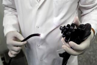
Instead, Chang said, he threads a scope tipped with an ultrasound sensor into the tract through the esophagus or rectum. The endoscope emits high-frequency sound waves that come back as echoes. The echoes are converted into electronic signals which, when portrayed on a television monitor in shades of gray, can reveal to the trained eye the presence of a tumor or enlarged lymph node.
After locating a lesion, which may be lurking in any of the five paper-thin layers of the gastrointestinal tract, Chang said, he uses a needle to make quick and painless biopsies that are sent to the laboratory for a more accurate diagnosis.
From the tract, Chang said, he also has easy access to parts of the lungs that are unreachable by other diagnostic techniques short of surgery.
The entire ultrasound and biopsy procedure takes up to two hours and can be done on an outpatient basis without general anesthesia. It costs $1,500 to $2,000, compared to $15,000 to $20,000 for exploratory surgery.
One of Chang’s patients, George Cornett, 67, of Buena Park, was prepared for surgery that would take six hours and involve cutting out most of his cancerous esophagus before Chang lowered an ultrasound endoscope down his throat and found that the cancer already had spread to a distant lymph node close to his stomach.
“It saved me a lot of grief, I believe,” Cornett said.
While endoscopic ultrasound has been available for five years in the United States, Chang said, it has been possible for U.S. physicians to use this technology to simultaneously image and sample cancerous tissue inside the body for about a year.
That is when Pentax Precision Instruments Corp. developed a new ultrasound endoscope that allows a physician to guide a biopsy needle with great precision into the masses that he finds. The new technology, Chang said, allows lymph nodes to be safely biopsied only a few millimeters from blood vessels.
From February, 1993, to February of this year, he said, he performed the new diagnostic procedure on 38 patients, with a mean age of 60, at UCI Medical Center and Long Beach Veterans Administration Medical Center.
He said he was able to diagnose cancer in 66% of the patients who previously were undiagnosed and discovered that 44% of the cancers were more advanced than other diagnostic procedures could have determined.
Perhaps most significantly, he said, the findings directly influenced the decision not to perform surgery on six patients, representing 26% of the patients with a previous cancer diagnosis, whose cancer had spread too far for surgery to help.
Not all information gleaned from the combined ultrasound and biopsy procedure is necessarily bad, said Chang. “In one instance a guy had a tumor in the pancreas and was scheduled for surgery. But the aspiration showed the tumor was not malignant, and so the surgery was canceled.”
Chang cautioned that the technology is not 100% accurate. In his research, about 9% of cancers initially were missed by the technology because the tissues that he sampled were not malignant.
Besides identifying inoperable cancers, the new technology also may be used in the future to make much earlier, life-saving diagnoses of cancers of the pancreas, Chang said. He said the technology may provide a non-surgical means for confirming the results of blood tests that are being developed to detect tiny cancers of the pancreas while they are still operable.
Diagnosing Inoperable Cancer
A new non-surgical technique developed by a UC Irvine physician combines ultrasound and a needle to determine when cancer is inoperable. The outpatient procedure decreases the need for exploratory surgery, takes 1 1/2 to 2 hours and does not require a general anesthetic. 1. Scope inserted down patient’s throat to stomach 2. Surgeon studies fiber optic and ultrasound images on TV monitors; uses hand-held device to maneuver scope left or right; follows needle’s movements on monitors. 3. If tumor is detected, surgeon protracts needle, inserts it into tumor. If needed, surgeon can insert a syringe through hollow needle to suction out a piece of tumor. 4. Surgeon examines tumor under microscope, and within 45 minutes can determine if it’s malignant. Scope: Three feet long; thickness of a small finger Ultrasound tip: Emits high-frequency sound waves that come back as echoes to TV monitor Rubber balloon: Filled with water to help ultrasound conductivity Fiber-optic lens: Relays images to TV monitor Retractable, hollow needle: Used to pierce suspected malignant tumor Light Source: Dr. Kenneth J. Chang; Researched by CAROLINE LEMKE / Los Angeles Times


