NAVIGATING THE MIND
- Share via
Imagine an atlas that maps each rolling contour of the human brain, charts the shoals of emotion, surveys the mountains of memory and locates each oasis of awareness.
Such a Baedeker of the brain would be an invaluable research guide for anyone trying to navigate the wilderness of the human mind. It could direct doctors as they plan neurosurgery, help psychologists better diagnose mental illness and aid therapists rehabilitating stroke patients. It could even come in handy for teachers trying to help students overcome learning disabilities.
Collecting data from a dozen analytical tools, researchers at UCLA, Caltech, the Jet Propulsion Laboratory and other research centers are assembling just such a comprehensive, computerized atlas of the world encompassed by the brain’s convoluted tissues. They are mapping the brain as part of a multimillion-dollar federal project.
“The technology has gotten to the point where you can chart the brain in a cartographic sense--from structure to function to health and disease,” said Arthur W. Toga, co-director of UCLA’s brain mapping division. “This is moving so fast--the technology of taking pictures of the brain--that it is almost science fiction.”
The brain mappers hope to produce computerized databases that will allow a researcher to move through the brain from the most fundamental molecular and chemical levels to three-dimensional neural anatomy and on to higher levels of brain function such as language, vision, hearing, sensation, movement and memory.
The task, they say, is so complex that it dwarfs the effort to map the entire human genome--the 100,000 genes that make up our hereditary blueprint. It may take a decade or more to accomplish.
To achieve such scope, their complete, computerized atlas would require:
* Maps of the brain’s internal structure, its neurons and their connections.
* The distribution of chemicals that support neural metabolism and communication between brain cells.
* The electrical properties of cell transmission.
* Blood flow and protein synthesis.
* Areas related to the action of various medical compounds and drugs.
As a foundation for this composite portrait of the brain, UCLA researchers are creating a definitive catalog of brain tissue for the National Library of Medicine. Slice by microslice, they are dividing the brain of a 51-year-old woman who died of breast cancer into 3,000 layers, each thinner than a human hair and scanning them into a computer database where they can be easily retrieved and examined.
Researchers then will start to overlay that basic map of the brain’s physical anatomy with views from new imaging techniques that record the physical changes in the living brain brought on by mental activity.
By documenting the performance of the normal human brain, the techniques give researchers the ability to construct comprehensive maps that link mental activity and physiology to detailed brain structures.
But any attempt to create a standard chart of the brain’s territory first must overcome the enormous difference in size and shape among human brains.
Not only are no two brains physically alike, but even the simplest mental activities--like watching a moving dot--can involve slightly different areas in different people’s brains, imaging studies show. Indeed, a single brain can go through an extraordinary range of physical changes in a lifetime, in response to learning, experience or disease.
Advanced computer graphics can help researchers compensate for such differences by allowing the images to be “morphed”--stretched or adjusted precisely into a standard form.
To better study the structure of the cerebral cortex--which accounts for most higher mental processes--neurobiologist Martin C. Sereno at UC San Diego recently developed a computer graphics program that allows him to inflate the cortex like a balloon, revealing details of anatomy otherwise hidden.
But for such techniques to become useful as diagnostic aids, the mapping experts first must develop a standard reference grid--a kind of latitude and longitude for the brain--to ensure that they do not distort the image when they enlarge or reduce it.
They must also ensure that the reference points are the same for all human brains. Otherwise, brain experts trying to navigate this always-changing territory of tissue will simply lose their way.
“Unlike the Earth, there is no accepted form of navigation for the brain,” said John Mazziotta, co-director of the UCLA brain mapping division. “Our challenge is to build [a computer model of] an average brain that encompasses all the probabilities of the structure.
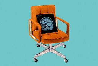
To build their atlas, scientists are relying on six different ways of taking pictures of the living brain:
* Magnetic resonance imaging (MRI) reveals the living brain’s structure with unprecedented clarity. MRI scanners use a powerful magnetic field to align atomic nuclei in the brain, then probe them with radio pulses to reveal concentrations of certain elements like hydrogen.
* Positron emission tomography (PET) shows how the brain uses energy, allowing researchers to track mental activity. PET scanners use radio activity to measure blood, glucose, or neurotransmitters such as dopamine as they move through the brain.
* Computerized arrays of electrodes (EEG and MEG) can track the electrical and magnetic signals underlying brain activity. MEG, short for magneto-encephalography, uses super-cooled, liquid-helium superconducting sensors to pick up the faint magnetic fields generated by active networks of nerves. EEG, which stands for electro-encephalography, records the much stronger electrical fields generated by the brain.
* Functional MRI (fMRI) allows researchers to monitor extremely high-speed changes in the blood flow through neural circuits as a way to pinpoint areas activated by mental activity. It also can detect the chemicals released by active neural cells.
* And the newest--a variation on PET scanning developed at UCLA--may eventually give researchers the ability to watch genes as they activate inside the living brain. The technique uses radioactive probes to home in on cells that contain a special “reporter” gene introduced by researchers. When that gene--and any other gene associated with it--is activated, the cells register on a PET scan as a bright spot.
Other scientists are using spectrometers to analyze the neurotransmitters used as chemical agents of communication between brain cells. At Caltech, they are using MRI microscopes to discover previously hidden anatomical details of neural cell structure.
Said Scott E. Fraser at Caltech’s Human Brain Project: “Each one of these things chips away at our ignorance.”


