Technology expands breast cancer screening options
- Share via
Breast-cancer-screening isn’t like looking for a needle in a haystack. It’s harder. It’s like looking for needles in a big field of haystacks, where some of the haystacks have needles, while most don’t, but you don’t know which are which, so you have to look in all of them.
Mammography is the best technique available right now to look for breast cancers in women who don’t have any symptoms. On average, screening mammograms correctly identify 80% to 85% of women who have cancer and about 90% of women who don’t.
Even as debate continues about who should get mammograms — and when, and how often — researchers are working on a blizzard of new approaches to breast imaging in hopes of reducing the number of cancers that are missed and the number of false alarms that lead to unnecessary biopsies. Here’s a look at some of the approaches under study.
Digital mammography
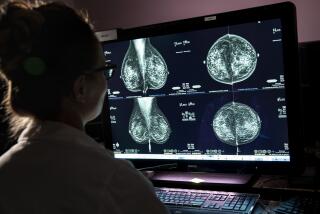
The screening mammography we get today takes X-ray images — usually two of each breast — looking for abnormalities that can’t be felt in a physical exam. These include small tumors and tiny deposits of calcium, called microcalcifications, which, in clusters, may be a sign of cancer.
It’s harder for mammograms to find cancers in the dense breast tissue that is normal in young, premenopausal women, because dense tissue and cancerous tissue each look white on a mammogram. Fatty breast tissue — more common in older women — looks dark on a mammogram, so any white cancerous tissue stands out.
In digital mammography, which was approved by the Food and Drug Administration in 2000, images are stored in a computer instead of on film. The technique may provide an edge in some cases.
A large clinical trial — the Digital Mammographic Imaging Screening Trial (DMIST) — compared the accuracy of conventional and digital mammography on nearly 50,000 women. Results, published in 2005, showed that across the entire sample, accuracy of the two kinds of mammography was similar, but digital was more accurate in premenopausal women, women younger than 50, and women with dense breasts.
Another study published last year found that breast cancer detection rates nearly doubled — from 4.1-4.5 per 1,000 women to 7.9 per 1,000 — at a diagnostic center in San Luis Obispo after the center switched to digital mammography.
Digital mammography has advantages: Images can be enhanced, and they’re easier to store and transmit. An analysis of 5,000 DMIST participants found that the average breast radiation dose per view was 22% lower for digital mammography than for analog.
But digital mammography is not available everywhere. “And it’s more important simply to get a mammogram than to wait to get a digital one,” says Dr. Pulin Sheth, chief of breast MRI and assistant professor of radiology at USC’s Keck School of Medicine.
Computer-aided detection
When it comes to reading mammograms, evidence shows that two radiologists are better than one — improving detection rates by about 10% on average. Double-reading is standard practice in many European countries but is used only in 25% to 30% of U.S. readings.
Could a computer fill in for the second reader? In this approach, a radiologist reviews the mammogram. Then a detection device scans it, marking suspicious areas. The radiologist then compares his or her analysis with the computer’s and decides if further evaluation is needed.
Research hasn’t always found benefits from computer-aided detection, but a large clinical trial published in 2008 did. Among more than 31,000 women who had conventional mammograms in England, computer-aided detection identified 87.2% of breast cancers, pretty much on a par with double-radiologist readings.
Stochastic resonance
Scientists have discovered that by adding the right kind of noise (interference) to a mammogram image, they can make the image clearer. In a study of 75 images published last year, researchers found they could detect cancers as well or better than mammography alone, while reducing the number of false alarms by as much as 36%.
Digital tomosynthesis
First reported in 2007, digital tomosynthesis takes at least 11 X-ray images of the breast at different angles, which a computer combines into three-dimensional images.
While mammography pulls the breast away from the body and squeezes it between two glass plates, tomosynthesis uses just enough pressure to hold the breast still — so it doesn’t hurt the way mammography can. And because it doesn’t squeeze the breast, tomosynthesis doesn’t create overlaps in tissue that hide cancers.
It also may result in fewer false alarms. In a study reported last year, radiologists interpreted images from 125 women, 35 who had cancer and 90 who did not. False-alarm recall rates were reduced by 30% by using both tomosynthesis and digital mammography instead of digital mammography alone.
Stereoscopic mammography
In this emerging technology, radiologists take two digital X-ray images of the breast about eight degrees apart and then fuse them into one stereoscopic view. At a 2007 meeting, researchers reported interim results for 1,093 patients with above-average breast cancer risk. Of 109 cancers, standard mammography missed 40, while stereoscopic missed 24 — 40% fewer.
Also, stereoscopic yielded 49% fewer false alarms — 53 compared with 103 for standard mammography — and was better at spotting microcalcifications.
Ultrasound (sonography)
In an ultrasound test, high-frequency sound waves travel through the breast, bouncing off boundaries — between fluid and tissue, for example, or between normal tissue and a tumor — and produce an image called a sonogram.
Ultrasound can distinguish solid tumors from fluid-filled cysts, but it’s not useful for routine screening because it misses too many cancers, including microcalcifications, and comes up with too many false alarms. Still, it’s sometimes used for screening patients with dense breasts (generally younger women), usually in addition to mammography.
Researchers are trying to beef up the technology.
One way is via elastography. Many ultrasound machines are equipped with software that measures the stiffness of a lump, capitalizing on the fact that malignant tumors are generally less elastic than normal, healthy breast tissue. In theory, such technology should help to distinguish malignant from benign tumors, and evidence is accumulating that it does.
In one study, presented at a meeting last year, researchers pitted elastography against regular ultrasound in 99 patients with a total of 110 breast tumors. Biopsies determined that 26 of the tumors were malignant.
Regular ultrasound correctly identified 23 of the 26 — but elastography correctly identified all 26.
Regular ultrasound had many false alarms, classifying 60 of the 84 benign tumors as malignant, while elastography had only 26 — less than half as many.
Another study found that following up mammograms with elastograms could reduce the number of biopsies by almost half, while nearly doubling the percentage of biopsies that found actual cancers.
Researchers are also working on creating three-dimensional ultrasound images and on distinguishing between benign and malignant breast lumps by, for example, using ultrasound to measure how fast blood moves through the lumps, with faster speeds associated with malignancies.
Magnetic resonance imaging (MRI)
Relying on the properties of magnetic fields, radio waves and the billions of hydrogen atoms in the human body, MRI produces precise images of internal organs and tissues based on differences in water content and distribution. A breast MRI produces hundreds of images from multiple directions.
MRI is not used for routine breast cancer screening because it’s expensive and tends to give a lot of false alarms, although the rate of these is decreasing, says Dr. Lawrence Bassett, the Iris Cantor professor of breast imaging at UCLA’s Jonsson Comprehensive Cancer Center.
An upside? MRI can detect cancers when they’re very small. “It’s almost at the point of being a screening tool for very high risk patients, to supplement mammography,” says Dr. Tova Koenigsberg, division chief of breast imaging at Montefiore Medical Center in New York City.
One problem might be acceptance. In a study of 1,200 women at intermediate or high risk for breast cancer, only 52% ended up completing the exam. The most common reason for turning down the offer was claustrophobia. A breast MRI usually takes about an hour, and the patient must lie very still inside a large tube.
Although MRI is very good at finding small lumps in the breast, it’s less good at determining whether the lumps are cancerous or not. Recently researchers have been working to mitigate this problem by using a procedure that can measure the cellular density of lumps. (Denser lumps are more likely to be malignant.)
They are also looking at ways to reduce MRI false alarms using proton magnetic resonance spectroscopy, a procedure that adds about 10 minutes to a standard MRI and provides information about the chemistry of a tumor. And some scientists are looking for better statistical ways to interpret the results of a breast MRI in order to reduce the number of false positives.
Electrical impedance scanning (EIS)
Electrical impedance — a measure of how fast electricity travels — can be used to locate breast cancers because malignant breast tissue conducts electricity better than normal breast tissue. Not currently approved for use as a screening device, it is approved for follow-up use when doctors aren’t sure from mammograms if a biopsy should be done. Some studies have suggested that electrical impedance scanning may be useful for identifying young, premenopausal women at increased risk for breast cancer.
Scintimammography (molecular breast imaging)
In scintimammography, a radioactive tracer is injected into the patient, then a special camera locates where the radiation accumulates in the breasts. It is not used as a screening test but has proved a useful complement to mammography in diagnosing cancers when they’re large enough. Some studies suggest that a newly developed “high resolution dedicated breast camera” may be useful with smaller cancers too.
The future
A note of caution: Although some of these new technologies are already in use and others are in clinical trials, most probably won’t be coming to a clinic near you very soon. And none of them offers hope for solving one big screening dilemma: how to distinguish dangerous cancers from those that will never go on to threaten a woman’s life or cause her any problems at all. Because scientists can’t do this yet, they generally treat all cancers, often with toxic chemicals or radiation.
Nonetheless, breast cancer screening has been shown to save lives, and scientists around the world are working to make it do an even better job. “This is a very active field of research,” Sheth says. “We keep learning more every day.”


