A Perinatology Glossary : ULTRASOUND
- Share via
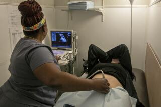
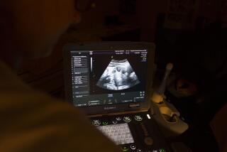
An imaging process that sends inaudible high-frequency sound waves through a transducer, a small device that the doctor moves slowly atop a pregnant woman’s abdomen. The sound waves are bounced off fetal tissue and organs, translated by computer and then projected on a video screen, creating a blurred, translucent image of the fetus. The use of ultrasound is a key procedure in prenatal diagnosis as well as in selective termination of pregnancy.
AMNIOCENTESIS
During the second trimester--usually at 16 to 18 weeks, but earlier in some cases--a doctor inserts a needle into the amniotic fluid that surrounds the fetus and withdraws fluid. Fetal cells floating in the fluid can be grown in the laboratory and analyzed to detect evidence of genetic defects and to determine the fetus’ sex. Results are available in about two weeks. Studies indicate that the accuracy of the test is 99.4%, while the risk of miscarriage or infection from the diagnosis is .2% or .3%. Most doctors do not routinely offer amniocentesis to patients younger than 35, unless they are at risk of having a child with a birth defect. After age 35, the risk of a woman’s having a child with Down’s syndrome, a genetic defect that leads to mental retardation, rises substantially. But many doctors consider “maternal anxiety” sufficient reason to offer the test to younger women.
CHORIONIC VILLI SAMPLING
A newer, experimental method of prenatal diagnosis performed during the first trimester,in which doctors use a catheter to pull cells from chorionic villi tissue, which will eventually become the placenta. Doctors then grow and analyze the cells to check for genetic problems or determine the fetus’ sex. The advantage of this technique over amniocentesis is that it can be performed earlier in pregnancy, enabling women to undergo earlier and less traumatic abortions. But the risk of miscarriage is slightly higher from this test than amniocentesis, and this procedure is somewhatless accurate.
SELECTIVE TERMINATION
In the first trimester of a pregnancy with multiple gestations--such as twins, triplets or quadruplets--a doctor reduces the number of fetuses in the uterus by guiding a needle through the pregnant woman’s abdomen and into the chest cavity of one of the fetuses. A lethal dose of potassium chloride is injected into the fetus, whose remains are absorbed by the mother. Still considered an experimental procedure, this is the most high-tech version of abortion. The widespread use of fertility drugs and in vitro fertilization has increased the number of multiple gestations. Selective termination is being developed because adverse outcomes in such pregnancies are directly proportional to the number of fetuses in the uterus, mostly because of a greater risk of prematurity.


