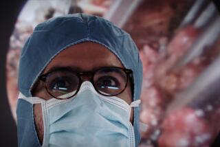
3-D, Computerized Cadaver on the Internet : Medicine: The ‘Visible Man’ is a detailed atlas of the killer’s body, assembled digitally from thousands of X-ray, magnetic and photo images of cross-sections.
- Share via
CHICAGO — Sixteen months ago, a killer was executed in Texas. Today, his body is a teaching tool for the world, made available on the Internet as the first three-dimensional, computerized cadaver.
The “Visible Man” is a detailed atlas of the human body, assembled digitally from thousands of X-ray, magnetic and photo images of cross-sections of the body.
The National Library of Medicine unveiled the “Visible Man” in late November at the annual meeting of the Radiological Society of North America.
“This is the first time such detailed information about an entire human body has been compiled,” said Dr. Donald A. B. Lindberg, director of the library, which is the equivalent of the Library of Congress for medical matters.
The digitalized cadaver will be available free to anyone who gets permission from the library. But the data is so extensive that downloading it takes up to two weeks of uninterrupted time on the Internet, and up to 15 gigabytes of storage space, enough to accommodate about 50 times the contents of The Encyclopedia Britannica.
The information would fill more than 30 typical personal computers and is expected to be sought mainly by medical schools and researchers, said Michael Ackerman, a computer specialist with the library.
The “Visible Man” will be an immediate teaching tool for medical students, and in the future, it could be used to develop surgery simulators much like the flight simulators used to train pilots today, he said.
“We hold this out as an example of the future of health care . . . which more and more will become visual rather than textual,” Ackerman said. “It’s a whole different way of looking at medicine.”
The library is spending $1.4 million to develop the “Visible Man” and a “Visible Woman,” which is still more than a year away, said Victor Spitzer, a computer-imager and anatomist at the University of Colorado Health Sciences Center in Denver, where the imaging was done.
The work began on Aug. 5, 1993, several hours after the execution by injection of Joseph Paul Jernigan, 39, an ex-mechanic who killed a 75-year-old man during a burglary and who left his body to science. The body was flown to Colorado and underwent hours of CAT and MRI scans.
Then it was sawed into four pieces and each was frozen in gelatin. One at a time, each piece was attached to a special table and slowly raised under a special planing tool called a cryomacrotome.
The instrument, designed especially for cutting cadavers, shaved away cross-sections of the cadaver 1 millimeter thick--a total of 1,870 cross-sections from head to toe. Each newly exposed layer of cadaver was photographed and scanned into a computer by a digital camera.
More to Read
Sign up for Essential California
The most important California stories and recommendations in your inbox every morning.
You may occasionally receive promotional content from the Los Angeles Times.


