Study finds flaws in computer-aided mammography
- Share via
An increasingly popular technology that uses computers to scan mammograms actually produces worse results than human reviewers using their eyes and experience, according to a study released Wednesday.
Radiologists using computer-assisted detection software were more likely to interpret a benign growth as potentially cancerous, researchers said in the New England Journal of Medicine.
The false-positive readings led to additional scans and needless biopsies, adding $550 million to the annual cost of breast cancer screening in the U.S., researchers said.
In addition, the computer-aided detection system, known as CAD, did not help radiologists find more real cancers, the report says.
Dr. Ferris Hall, a radiologist at Beth Israel Deaconess Medical Center in Boston, who wrote an editorial accompanying the report, said some mistakes might have been the result of inexperience with CAD. It takes radiologists several years to learn the technology, he said.
Nonetheless, Hall said, the study is a setback for the technology, which is used in 30% of the more than 30 million mammograms performed in the U.S. annually. Many imaging centers promote their CAD services, which are potentially more profitable than standard mammography, he said. Medicare pays an additional $20 for each mammogram screened by CAD.
“This will have a major impact on radiology,” Hall said. “They were calling people back for more scans and did more biopsies -- that is hurting people. And what did they get for it? No significant increase in cancer detection.”
The study shows other techniques are needed to detect breast cancer in its earliest stages, said Dr. John E. Niederhuber, director of the National Cancer Institute, which funded the study.
Breast cancer is the second most common cancer in women, next to skin cancer. The American Cancer Society estimates that 178,480 women will be diagnosed with the disease this year and that 40,460 will die of it.
Mammography, an X-ray image of the breast, has long been the primary tool for detecting breast cancer in its earliest stages, before tumors are large enough to detect in a clinical breast exam.
Last week, the American Cancer Society recommended annual magnetic resonance imaging for women at high risk for breast cancer.
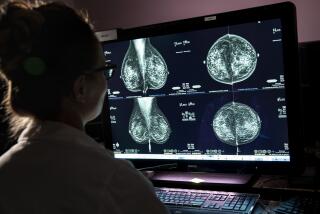
Launched in 1998, CAD was designed to improve the accuracy of mammogram readings. A device converts X-ray film into a digital file that can be analyzed by computer and displayed on a monitor. The software marks suspicious areas on the screen image for the radiologist to review in addition to the areas detected by the radiologist’s eyes.
Previous studies assessing the benefits of the technology have produced mixed results.
The latest study looked at mammography results for 220,000 women at 43 imaging centers in Colorado, New Hampshire and Washington state. Seven of the centers began using CAD during the study.
The researchers found that after using the software, the rate of women recalled for additional imaging tests went up 32%, along with a 20% increase in the number of breast biopsies. The women were later found not to have breast cancer.
Maryellen Lissak Giger, professor of radiology at the University of Chicago and an inventor of CAD technology, said the study had several flaws. It was too small to detect a slight improvement in cancer detection, she said. Moreover, the research, from 1998 to 2002, evaluated an early version of the CAD system, said Giger, who owns stock in Hologic Inc. of Bedford, Mass., which licenses its technology from the University of Chicago.
*


