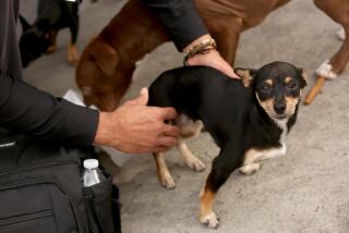
High-Tech Medicine Goes to Dogs--and Cats : Medicine: Fido and Fluffy can get the same diagnostic treatment as their owners in an Auburn University veterinary clinic.
- Share via
AUBURN, Ala. — Gone are the days when a stethoscope, an X-ray machine and educated guesswork were a veterinarian’s best tools for diagnosing an animal’s ailment.
Veterinary medicine has entered the high-tech age, and a clinic at Auburn University is giving dogs and cats access to some of the glitziest gadgetry available for either people or pets.
Two devices at the $1-million Ware Diagnostic Imaging Center allow vets to look inside animals without picking up a scalpel. The combination of a magnetic resonance imaging machine and CT scan equipment on a veterinary school campus is a first in North America, Auburn officials say.
“State-of-the-art equipment, needless to say, is not the bailiwick of the average veterinarian’s office,” said Dolores Jenkins, a spokeswoman with the American Veterinary Medical Assn.
John T. Hathcock, a radiology professor at Auburn, said the use of MRI and CT scanning is a big step for vets, especially since their patients can’t say where it hurts.
“One of the emphases will be on diagnosing and researching cancer,” Hathcock said in a recent interview.
“With a regular X-ray, all the body parts are superimposed on each other,” said Hathcock. But with MRI and the CT scanner, he said, the body can be seen in sections, “like slices in a loaf of bread.”
CT--or computerized tomography--scanning has been around since the early 1970s. The procedure involves placing a patient on a movable table surrounded by a huge, doughnut-shaped X-ray machine. As the table moves, the machine takes X-rays that show a cross-section of the body. Tumors and other ailments show up on a video screen.
The Auburn center has been performing CT scans on animals since late October, for $200 to $250 each, Hathcock said. The patients come primarily from veterinarians who refer tough cases to the school.
Across the hall from the CT scanner is the magnetic resonance imager, which looks more like a Greek temple than a piece of medical equipment.
A patient is placed on a movable platform. The animal is centered under a table-like structure, with four thick, round legs. A huge magnet inside the “table top” aligns hydrogen protons in the body, and an image is produced by monitoring the resulting atomic movement.
While CT scanning produces a good picture of internal structures, MRI is sharper, with more anatomy revealed.
Both machines are set up for small animals, Hathcock said, but the radiology department plans to adapt them for use with larger animals. “If we could do a leg of a horse it would be a great help. With horses, that’s where most of the injuries are,” he said.
Bill Adams, a veterinary radiologist at the University of Tennessee in Knoxville, said most vets have some catching up to do. Tennessee’s veterinary school has access to a CT scanner and other imaging equipment, Adams said, but Auburn is alone in having the machines on campus.
“Because of economic concerns, (veterinarians) have been a few steps behind our human counterparts,” he said.


