Focus on Pregnancy : The Pregnant Pause : With more choices in prenatal testing, women and their doctors must discuss and carefully consider which test is right for them.
- Share via
When Carrie Yoshida was born in 1957, the doctor who delivered her looked at the newborn’s deformed left hand, shrugged and told her startled parents: “A birth defect.”
That was the extent of the explanation for Carrie’s hand, in which several fingers were fused together and resembled a cat’s paw. After a series of plastic surgeries to separate the fingers, Yoshida went on to play volleyball and teach art.
But when the Redondo Beach woman became pregnant at age 41 last year, the “birth defect” explanation seemed wholly inadequate. Carrie and her husband, Wendall, wondered if the problem could be passed on to their baby.
After providing a genetic counselor with an extensive health history, Carrie underwent a procedure known as chorionic villus sampling (CVS), in which a small piece of tissue from the placenta is removed via needle to examine the baby’s genes and look for genetic diseases. And midway through her pregnancy, Carrie had an ultrasound, which clearly showed a baby with 10 perfect fingers and toes.
“If my child had [the hand defect], I felt that wasn’t a reason to terminate the pregnancy. But I wondered if the child might have another problem” related to the hand abnormality, says Carrie, who gave birth to a son, Cole, on July 29. “I think I would have been very stressed if I had not found out.”
Clearly, the Yoshidas benefited from appropriate use of prenatal testing. But, after a decade of advances in prenatal screening and genetic testing, doctors are not always in agreement about which tests are best suited for particular patients.
“The only people who are certain about what should or shouldn’t be offered are the plaintiffs’ lawyers,” Detroit obstetrician Marc Evans says wryly, noting the frequent lawsuits that are filed by parents whose baby is born with an abnormality that they think should have been detected during pregnancy.
According to Evans, a leading authority in prenatal testing and a professor of genetics at Wayne State University in Detroit, the field of prenatal testing is shifting daily, with new advancements on the horizon and once-popular practices falling out of favor. These changes mean that pregnant women and their partners no longer can depend on the advice of friends who gave birth three or four years ago.
Couples need to do their homework and have detailed discussions with their doctors or other health care professionals about their options for prenatal testing, experts advise. Here is an update on the major tests and issues surrounding them.
Ultrasound
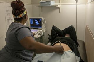
In a darkened, closet-sized room in the offices of Dr. Lawrence Platt at Cedars-Sinai Medical Center in Los Angeles, a video monitor displays the three-dimensional image of a fully formed fetus seemingly floating in space.
The astonishingly clear images look as if they were taken with a video camera tucked in the mother’s uterus. To an untrained eye, the baby looks as it should. But, in fact, there are problems, Platt says.
The fingers are too short and chubby. The chin is pulled back into the face. The forehead is very flat. The fetus, in fact, has a major chromosomal malady called trisomy 18, which will likely result in death shortly after birth.
The video of this baby illustrates both the promise and drawbacks of the latest sensation in prenatal testing, 3-D ultrasound. By viewing the fetus in three dimensions, doctors can more accurately discover or confirm problems. However, expectant parents may find it extremely hard to reconcile the exquisite pictures with the knowledge that this baby will be born gravely ill.
“It’s a great advance in technology. But it shouldn’t be abused. We’re still learning the power of this technology,” says Platt, chairman of obstetrics and gynecology at Cedars and a leading researcher in ultrasound technology.
Ultrasound technology creates pictures of the uterus and baby with sound waves. The test is considered very safe, although studies have never established with certainty that the procedure is not without risk for the mother or baby.
With 3-D ultrasound, the test is performed the same way as conventional ultrasound, except that the computer gathers images from a third angle. Thus, a 3-D picture creates a feeling of depth and allows the doctor to view the fetus from different angles. In contrast, 2-D ultrasound requires the doctor to use his or her imagination to complete a picture of the baby.
The advanced ultrasound technique--whose $500 cost is usually covered by health insurance if the procedure is “medically necessary”--is not intended for use in all pregnancies, doctors say. Studies have shown that the procedure is superior in diagnosing abnormalities of the face, brain, fingers and feet, skeleton and lower extremities.
The technology also requires experienced operators, Platt says. Typically, only large, academic medical centers in the United States have 3-D ultrasound and are using it only in cases where an abnormality has already been detected.
Eventually, the technology may become most popular in a screening test for Down syndrome called nuchal translucency. Nuchal translucency is an area of fluid accumulation between the skin and subcutaneous fat at the nape of the neck and is a possible indicator of Down syndrome.
With 3-D ultrasound, researchers may be able to come up with an exact measurement of nuchal translucency so that a computer receiving the data could determine the probability of Down syndrome, Platt says.
But this new procedure already is raising thorny ethical issues for parents who must decide what to do when the test results come with bad news. With 3-D ultrasound comes the fear that detailed information early in the pregnancy will lead couples to terminate pregnancies in which there is a correctable and nonlife-threatening disorder, such as webbed fingers or a cleft lip.
A study of Israeli women, published in March in the Cleft Palate-Craniofacial Journal, found that 23 out of 24 women who learned during their 15th week of pregnancy that their babies had cleft lip decided to have abortions.
In a similar study in San Diego, two of eight women terminated their pregnancies. Unlike their Israeli counterparts, however, the San Diego women didn’t learn about the cleft lip diagnosis until their 24th week--or sixth month--of pregnancy, after which most doctors will not perform an abortion.
Amy Mackin, hotline manager for the Cleft Palate Assn. in Chapel Hill, N.C., says she is seeing an increase in calls from couples who have received a prenatal diagnosis indicating cleft palate.
“Getting the diagnosis prenatally gives them more time to prepare. They can have the special bottles they’ll need and a surgeon lined up. I think [prenatal diagnosis] is good in a lot of ways. But there are also these ethical issues,” she says.
Platt acknowledges that use of 3-D ultrasound early in the pregnancy may lead more couples to terminate pregnancies in which there is a correctable birth defect.
“I don’t feel that this technology is search-and-destroy. But people are going to use this to whatever means they wish. . . . And it’s a patient’s right,” Platt says.
Some women with normal pregnancies are also getting 3-D ultrasounds, although this use of the technology is frowned on by some medical researchers.
A few centers have opened around the country offering 3-D pictures to pregnant women willing to pay $500. Dr. Robert Z. Gergely opened the 3-D Sonography Center of Beverly Hills to give “peace of mind” to expectant mothers.
“Invariably, no matter what we tell the mother about her baby’s health, she says, ‘Yes, but I want to see.’ If I can accomplish that three or four months before her due date and lower her anxiety, that accomplishes a lot,” Gergely says.
Because Gergely offers the 3-D pictures at about 28 weeks of pregnancy, he says that most of the babies with abnormalities have already been detected through earlier tests. However, he says, problems spotted during the ultrasound are reported to the patient’s doctor.
In such cases, the expensive procedure is not covered by insurance because of the lack of medical necessity.
Critics of this practice, however, say it’s a misuse of technology because ultrasound has not been proved to be completely safe and because couples might believe that beautiful pictures mean the baby is fine--a guarantee no one can make.
“I have grave concerns about it,” Platt says. “It’s the ‘wow’ effect. People should not expect this as a routine part of prenatal care.”
Alpha-Fetoprotein, Multiple-Marker Tests
Screening tests are different from diagnostic tests like amniocentesis and CVS, because they only indicate a possible problem and carry no risk to the mother or baby. Thus, screening tests are offered to all pregnant women.
The most popular screening test is a blood test that checks for alpha-fetoprotein, a protein produced by the fetus. AFP can detect between 60% to 80% of abnormalities, often neural tube defects, such as spina bifida. Positive tests must be verified by amniocentesis or CVS.
Researchers are also looking at other “markers” in a woman’s blood that can indicate the presence of Down syndrome. While tests looking for three substances (AFP, human chorionic gonadotropin and estriol) are already in use, studies are underway to add one or more other markers to the list.
“All these markers are indications of Down syndrome. We hope that adding them further increases the accuracy of the test and reduces the number of people who need invasive procedures,” Evans says. “Even some women over 35, in the face of having a reassuring screening test, are choosing not to have invasive testing.”
Curbing the rate of invasive testing could result in big savings, Evans says.
“If you can reduce the number of people undergoing amniocentesis from 6% to 5% [of all pregnant women], you could save $70 million a year in this country,” he says.
Amniocentesis
The mainstay of prenatal genetic testing, amniocentesis carries a slightly higher risk of miscarriage--about a half-percent beyond normal. Earlier this decade, there was enthusiasm for performing amniocentesis in the first trimester, around 11 to 14 weeks. Diagnoses made in the first trimester are preferable because a couple can learn of a problem before they have announced the pregnancy. And pregnancy terminations carry much lower risks than when performed later.
Fears of a higher rate of miscarriage have dimmed enthusiasm for “early” amniocentesis. Doctors who have performed a large number of these tests, however, typically don’t experience higher miscarriage rates in their patients, says Dr. John Williams III, co-director of reproductive genetics at Cedars.
Chorionic Villus Sampling
Like early amniocentesis, CVS started out with a bang of positive publicity because it can be performed in the first trimester. But enthusiasm for the test waned because of fears that it increases the risk of limb defects in babies.
Williams, who operated one of the largest prenatal testing centers in Los Angeles in the early ‘90s, performed about 1,000 CVS tests in 1990. By 1992, that number had dropped to about 600, and its popularity has continued to fade, he says.
However, Williams says, subsequent studies have not found an increased risk in limb defects. Medi-Cal--the state medical insurance program for low-income families--even began paying for CVS this year, suggesting the procedure is in no way considered high-risk or experimental.
“If people knew the correct information on CVS,” Williams says, “we’d see double or triple the numbers we’re seeing now.”
(BEGIN TEXT OF INFOBOX / INFOGRAPHIC)
Choices in Prenatal testing
Some prenatal tests are used to detect the possiblility of a problem. Such tests are low-risk and are usually offered to all pregnant women. Diagnostic tests are typically invasive procedures and recommended for women at higher risk of experiencing an abnormality.
SCREENING TESTS
Alpha-fetoprotein
15-18 weeks
Mother’s blood sample. Screens for high risk of neural tube defects, such as spina biffada, and a few other abnormalities.
Multiple marker
15-18 weeks
Mother’s blood sample. Screens for Down syndrome.
SCREENING / DIAGNOSIS TESTS
2-D Ultrasound Weeks
Typically 8-22 weeks; can be done any time
Uses sound waves to create pictures. Can detect structural abnormalities and provide information about fetal growth.
3-D Ultrasound Weeks
8-28 weeks
Similar to 2-D but adds another plane to picture. Confirms suspected problem; detects structural abnormalities not seen in 2-D.
DIAGNOSTIC TESTS
Amniocentesis
15-20 weeks
Needle inserted to draw small sample of amniotic fluid and analyze fetus’ DNA.
Early amniocentesis
11-14 weeks
The same as amniocentesis but carries additional risks.
Chorionic villus sampling
10-12 weeks
Needle inserted to remove tiny piece of tissue from placenta and analyze DNA.


