Pill-Size Gastro-Cam Is Just a Swallow Away
- Share via
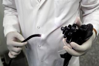
Having one’s innards examined for cancer or other irregularities with the use of long, flexible cables is nobody’s idea of fun. And viewing some parts of the gut involves particularly long lengths of cable and procedures that are awkward for the doctor and just plain nasty for the patient.
Now British and Israeli scientists report that they have developed something much more dignified. It’s a pill-size camera that is simply swallowed, and--as nature takes its course--inches down the gastrointestinal tract, taking snapshots of the stomach, small bowel and parts of the colon before being voided at the other end.
“It was easy to swallow--just a mouthful of water and it had gone,” said Dr. Paul Swain, gastroenterologist at the Royal London Hospital England, a coauthor of the report and the first person to swallow the device.
Being able to view images from his small bowel--the hard-to-reach 20 feet of gut that lie between the stomach and the colon--without pain and discomfort was “quite thrilling,” Swain said. “I’m one of the few people who has seen their [own] small bowel and smiled.”
The fantastic device, described last week in the journal Nature, is called a wireless capsule endoscope, and is one of several new technologies aimed at better charting the guts coiled within our bodies.
Researchers want to more easily view the small bowel, the length of tubing that lies between the stomach and colon, where bleeding from malformed blood vessels can sometimes cause anemia. And they are investigating new techniques for viewing the colon--site of the second most common human cancer--to supplement the conventional endoscope exam.
The immediate promise of the new, miniature camera, at least in its current form, lies in viewing the small bowel.
“It’s exciting,” said Dr. Michael Kimmey, professor of medicine at the University of Washington in Seattle, and president of the American Society for Gastrointestinal Endoscopy. “I think this will be a major way to improve diagnosis of small intestine problems, especially bleeding and benign tumors.”
Measuring just 1.2 by 0.4 inches and rigged out with a tiny camera, battery, transmitter and light source, the device is a marvel of compactness. Even five years ago, it would have been five to 10 times larger--more the size of a horse pill, said Arkady Glukhovsky, a coauthor of the Nature paper and a biomedical engineer with Given Imaging. Advances in imaging technology and chip circuitry made the shrinkage and six hours of battery life possible, while another development enables the essential white light illumination.
As the swallower goes about a normal day, the capsule completes its 30-foot journey in about 24 hours, pushed along by the natural squeezing action of the intestines. As the camera nudges ever onward, it rubs against the gut wall, which keeps the lens clean of debris so that clear pictures can be taken before the camera battery runs down.
No need to retrieve the camera once it passes out of the body. Images are transmitted continually to a detector worn on the patient’s belt, which the doctor later collects. Should an abnormality be detected, the doctor will know where in the gut it lies, based on the relative strength of signals transmitted to six separate aerials on the belt.
So far, the camera-pill, which was developed by engineers at Given Imaging Ltd., an Israeli company, in collaboration with Swain, has been tested on 10 people with normal insides. Now the inventors are seeking regulatory approval in several countries to study it in people with recurrent, unexplained gastrointestinal bleeding.
Some experts, though intrigued by the device, said its current usefulness is limited.
“The technology is fascinating,” said Dr. Ted Stein, director of endoscopy at Cedars-Sinai Medical Center. “But I think we need to wait for further developments.”
Stein and others point out that the device cannot be maneuvered like conventional endoscopes to reexamine areas of interest and cannot take a biopsy if a trouble spot is identified. Swain and colleagues argue, however, that diagnosing and locating the source of a small bowel problems is very valuable.
Also limiting, experts say, is the fact that the capsule looks at the small bowel but not the large--since it runs out of battery power before it explores more than a smidgen of the colon.
But it is handy for viewing structures that cause small bowel bleeding, called arteriovenous malformations. These tend to be flat and hard to view by a commonly used method, the barium X-ray, and very uncomfortable by the alternative method--threading long endoscopes through the nose then pushing them past the stomach into the small bowel.
But such cases are rare, doctors say. The small bowel is not often a site for disease. While about 100,000 people will be diagnosed with colon or rectal cancer in the United States this year, the number of small bowel cases will number in the hundreds, said Dr. Paul Engstrom, medical oncologist at Fox Chase Cancer Center in Philadelphia and consultant for the American Cancer Society.
Thus a much more useful technology, said Stein, would be one that could screen the colon for cancers and polyps. A traditional colonoscopy works well but is invasive, requiring a tube to be inserted through the rectum. As medicine moves toward recommending routine colon screens past the age of 50, and as it debates the best screening method, cheaper or less invasive first-order screens could prove important.
With this in mind, scientists have been developing so-called virtual colonoscopy, which uses X-ray imaging to give three-dimensional pictures of the bowel. The technique is not yet as accurate as regular colonoscopy, said Kimmey. It misses the smallest polyps, and blobs of fecal matter can be mistakenly scored as suspicious.
If the capsule camera could somehow be modified for the colon, said Stein, and if it were accurate and thorough, “it could potentially screen the population at large for colon polyps. If you could take 100 million people and say two-thirds don’t need a colonoscopy because they’ve had a capsule test, that would be very cost-saving, and very appealing.”
The capsule isn’t the only futuristic gut-exploring device under development. Other scientists are trying to develop robots that might one day slither through our innards seeking out and treating abnormalities.
Joel Burdick, a professor of mechanical engineering at Caltech, and Dr. Warren Grundfest, director of laser research and technology development at Cedars-Sinai Medical Center and a professor at UCLA, have been working on just such a robot. One day, they say, it could take photos and biopsies and perform surgery.
The snakelike prototype can wriggle down 10 feet of rubber hose and a couple feet of pig intestine in the lab. The team is reworking the prototype so that it can avoid damaging the small bowel’s fragile blood vessels and better manage the widely varying width, stretchability and slipperiness of the intestines.
“It’s going to be several years in development,” Grundfest said.
Gut Cam
A disposable, camera-equipped capsule may someday replace more invasive methods of examining the digestive tract. As the capsule moves naturally along the intestines, it illuminates its way with built-in lights and transmits video images to a small receiver worn by the patient. The system records a video clip up to six hours long, which is later downloaded to a computer and processed for viewing.
*
Source: Given Imaging


