Seeking a Clearer Picture
- Share via
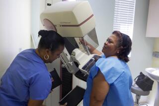
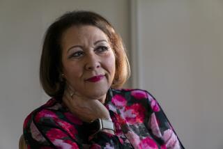
Judy Perotti didn’t think it was a big deal when she put off scheduling the mammogram that her doctor had ordered. When she finally had the exam about a month later, she was floored by the result: a suspicious mass, later determined to be cancer.
“I was only 44,” she says. “I was perfectly healthy, and I didn’t have a family history of cancer.”
Perotti, who underwent a lumpectomy and remains cancer-free 12 years later, believes the mammogram saved her life. “The cancer probably would have spread to my lymph nodes if I had waited another two years until it was large enough to feel,” she says.
Stories like Perotti’s have made mammograms the first line of defense against breast cancer, which is the leading cause of death among younger women, claiming 44,000 lives in the United States annually. Mammography can detect cancers in their earliest stages, giving patients a much better shot at long-term survival. And often the regular screenings can eliminate the need for debilitating chemotherapy, radiation and disfiguring surgery.
Today, about 30 million women get the exams annually. Yet questions about mammograms linger, especially among younger women who may be unsure about what steps to take to protect themselves. Women don’t know when they should schedule their first exam or how often to get tested. How much faith should they place in the results? Is there anything else they can do to up their odds of detecting tumors? And do these tests offer protection to women with a family history of breast cancer, whose risks of developing the disease are double that of their peers?
Some confusion stems from the fact that “mammography is not perfect,” says Dr. Lawrence W. Bassett, director of the UCLA breast imaging center. “We’re still missing 15% to 20% of cancers.”
And by the time a tumor is large enough to show up on a mammogram, it’s normally been growing for eight to 10 years. The failure to spot some tumors delays lifesaving treatment.
On the other hand, there’s a high rate of anxiety-provoking false positives, which means women have to get repeat tests, or invasive--and mostly unnecessary--biopsies to determine whether trouble is brewing inside their breasts.
The value of yearly mammograms for women in their 40s was a topic of debate among medical experts in the 1980s and early 1990s. Research at the time suggested that yearly mammograms offered little benefit to women in their 40s and missed about 25% of cancers in this age group.
The issue became an emotional flash point, and organizations such as the American Cancer Society, the National Cancer Institute and the American Medical Assn. advocated yearly screenings for women in their 40s, even though benefits weren’t clear.
But recent research seems to have settled that dispute. Newer studies, using more sophisticated techniques, demonstrated that annual mammograms trimmed breast cancer deaths by 25% to 36% among women between the ages of 40 and 49, which is comparable to rates among older women. And the exams have been proven to cut the death toll among women older than 50 by about one-third.
“The evidence now is pretty conclusive that yearly mammograms for women over 40 are beneficial,” says Dr. Edward Sickles, a professor of radiology at UC San Francisco.
Breast cancer is more difficult to diagnose in younger women because their breast tissue is thicker, or denser, which makes it harder to identify tumors (breast tissue usually thins as women age). And early-stage cancers may start as calcification spots that can be missed by routine screening.
Exacerbating the problem is that cancers in younger women tend to be more aggressive, which makes early detection even more crucial. In fact, one out of every five new cases of breast cancer, or 46,000 women, each year is diagnosed in women younger than 50. In most cases, there is no family history of the disease.
“Waiting two years between mammograms could be a death sentence for women with fast-growing tumors,” says Dr. D. David Dershaw, director of the breast imaging center at Memorial Sloan-Kettering Cancer Center in New York.
But mammograms aren’t infallible. Consequently, doctors urge women to get yearly manual breast checkups by a trained specialist, and do monthly self-exams to detect irregularities.
Kristin Reams, for example, got annual mammograms and had always received a clean bill of health. So when she noticed a lump in her breast while dressing one morning in 1995, she wasn’t alarmed. “I felt foolish calling the doctor,” recalls the 64-year-old Lancaster woman. “But I knew it was serious when they told me to come in right away.”
An ultrasound test, in which echoes from high-frequency sound waves form an internal picture, indicated a suspicious mass in one of Reams’ breasts. A follow-up biopsy confirmed that Reams had a cancerous tumor, about 2 centimeters in size. She had a mastectomy to remove her right breast but didn’t have chemotherapy or radiation. “I was very lucky, because they caught it early enough,” says Reams, who now unfailingly does monthly breast self-exams.
Given mammography’s limitations, medical scientists are striving to devise a breast cancer diagnostic tool that’s more accurate in identifying renegade cells before they turn deadly. There are several new techniques that show promise.
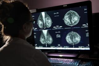
In January, the U.S. Food and Drug Administration approved digital mammography, in which a computer generates images with better contrast resolution than mammograms, enabling doctors to spot telltale little white flecks that indicate the presence of early stage cancers. The technology allows doctors to adjust the contrast or brightness, and to manipulate the image to improve clarity.
Right now, detection rates for digital mammograms are comparable to those of conventional mammography. As doctors become more proficient with the technology, however, digital mammography may supplant the conventional exam.
For now, UCSF’s Sickles says it is too soon to know if digital mammography--a far-costlier technology than standard mammography--will yield better detection rates over the long run.
Another new technology, called the ImageChecker, is designed to eliminate errors by doctors who “read” mammograms. Routine screenings miss 20% of cancers. Research shows that about half of these undetected tumors result from radiologists who are fatigued after looking at dozens of mammograms a day.
The ImageChecker is a computerized system that acts like another pair of eyes: It analyzes the image for regions suggestive of calcification clusters, tumor masses or distortions that are characteristic signs of cancer. Worrisome areas are then highlighted so the radiologist can review the mammogram. But the ImageChecker, which was approved by the FDA in 1998, is used at only a handful of sites across the country. An Internet-based company, iMammogram.com (https://www.iMammogram.com) of Westlake Village, is offering an ImageChecker service for $75.
However, UCLA’s Bassett cautions that the computer-aided detection device also picks up many irregularities that are not signs of cancer, and possibly leads some patients to undergo unnecessary biopsies.
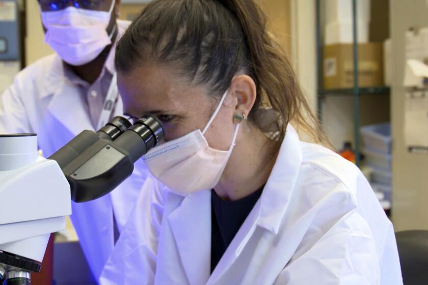
Another new test, called ductal lavage, holds out hope to high-risk women of detecting cancer at its earliest stages, up to a decade before tumors are large enough to be picked up by a mammogram. This procedure entails threading a thin tube through the nipple to suction out cells from the milk ducts inside the breasts. The cells are then examined in a laboratory for any abnormalities. Studies have shown that 95% of all breast cancers begin in the cells lining the milk ducts.
Preliminary results from a recent study of more than 500 high-risk women were encouraging. All of the women had normal mammograms and physical exams. But the tests revealed that there were atypical cells in 15% of the breasts studied, and suspicious or malignant cells in 5% of the women. “For the first time we have access to the sites where the vast majority of cancers start, which means that in the future we might be able to prevent cancers from developing,” says study co-author Dr. Laura J. Esserman, director of the Carol Buck Frank Breast Center at UCSF. Still, while these novel techniques are promising, they have not been subject to the same scrutiny as mammography, which has been studied exhaustively since the 1960s. “Basing any medical decisions solely on this new technology is premature,” says Dershaw, of Memorial Sloan-Kettering. “Mammography is still the best tool we have.”


