Testing for abnormal embryos
- Share via
Twenty-five years after in vitro fertilization gave birth to a new industry, there have been many improvements in conceiving babies in the lab. As the union of infertility treatment and genetic science, preimplantation genetic diagnosis may be one of the most far-reaching developments. PGD awaits further improvements that are considered key to whether it becomes the big thing or just a big thing in assisted reproduction. Here’s how it’s done:
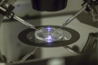
First, fertility doctors use drugs to stimulate a woman’s ovaries to produce many eggs and, after surgically removing them, transfer them to a lab, where a technician fertilizes them with a man’s sperm. The embryologist, a specialist in the earliest stages of fetal growth, cultivates the dividing “pre-embryo” in an ooze of growth-promoting gel.
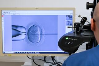
At day three, when the embryo reaches roughly eight cells, a microscopic cut is made in the embryo’s shell and a single cell (sometimes two) is sucked into a pipette. The cell is fixed to a slide and its DNA is made to replicate itself -- like multiple photocopies -- so its genetic make-up can be subjected to a series of probes. At this point, testing begins in a laboratory.
In PGD, a procedure for those at risk of certain known inheritable diseases, the geneticist goes directly to the genes most implicated in the disease and looks for duplications, unmatched or mismatched pairs, or fractured structures. Some families carry genetic “translocations,” where chromosomes are switched, and PGD now can check for these inheritances as well. Any irregularities on the affected chromosome could be evidence that the embryo would be affected by the familial disease.
Aneuploidy screening is a new adaptation of PGD. It identifies irregularities on a larger number of chromosomes and is conducted for women who have had several failed IVF cycles, miscarriages or who are nearing or older than 40. Once the embryonic cell is fixed and duplicated, fluorescent probes are inserted into the cell’s genetic material, three or four at a time. The probes attach themselves to particular chromosomes, color-coding pairs with the same hue. Because the most common genetic abnormalities tend to appear on a small handful of chromosomes (and because the range of display colors is limited by current technology), only seven pairs of chromosomes, plus the sex chromosomes, (the matched XX for a female, an unmatched X and Y for a male) are routinely tested in aneuploidy screening.
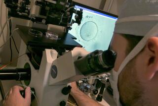
Like a host surveying a dance floor, the geneticist peering into the microscope starts counting, looking for unmatched chromosomes or for chromosomes with three copies rather than two. That aneuploidy -- or absence of match -- is highly common in the embryos of women older than 35; as many as 70% of the embryos that come from a 40-year-old woman undergoing IVF show evidence of this “spontaneous” (i.e., not hereditary) genetic abnormality.
For many years, embryologists have looked at an embryo’s external structure -- the number and shape of its cells and the smoothness of its skin -- to decide whether and which embryos should be transferred from petri dish to uterus. And a lot was riding on what some fertility specialists call “the beauty contest.” If a couple produces a number of embryos, doctors must decide which ones appear to have the best chance of success. If they put too many in a woman’s uterus, she could carry twins, triplets or even quadruplets -- with attending health risks and family disruption. Too few and she may not become pregnant. The wrong ones and the costly and painful IVF cycles leaves a couple baby-less.
PGD has shown fertility specialists that when it comes to judging an embryo, beauty matters but it’s what inside that counts. Many genetically abnormal embryos look perfect. But they will either not implant, result in a pregnancy that will end in miscarriage, or produce a child with a genetic abnormality like Down’s syndrome.
Guided by this information, fertility doctors can pick a relatively small number of genetically normal embryos -- the norm has become two -- to transfer to a woman’s uterus. By limiting the embryos transferred, they reckon they can virtually eliminate the likelihood of triplets or quadruplets and will likely drive down the rate of twins produced through IVF.
In screening the embryos produced by one woman in a single IVF cycle, geneticists such as Dr. Harvey Stern, head of the Genetics and IVF Institute’s PGD program in Fairfax, Va., say they see tremendous variations. “You’ll see some that are very, very abnormal, and then you’ll see one that is just stone-cold normal. You think, ‘Oh my God, how does anyone get pregnant?’ ”
And when the chromosomes light up in perfect, color-coded pairs, revealing an embryo free of apparent disease or obvious abnormality? “It looks good,” says Stern enthusiastically. “It looks really nice. It makes you feel like, this is really working.”
*
-- Melissa Healy


