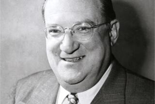
Henry Molaison’s brain lives on
- Share via
Reporting from Hartford, Conn. — Henry Molaison lived in relative obscurity, but he possessed one of the world’s most famous brains.
Known to generations of scientists and psychology students as H.M., Molaison lost the ability to form new memories after surgery removed part of his brain and, by agreeing to be studied over several decades, transformed the way we understand memory.
H.M. died last December, but science isn’t done with his brain.
Molaison, a Hartford, Conn., native who in life often expressed a wish to do what he could to help people, donated his brain for research. The brain now sits in the lab of neuroanatomist Jacopo Annese, director of the Brain Observatory at UC San Diego, where researchers have big plans for it.
Last week, a year after Molaison’s death, Annese and his team began cutting the brain into thousands of slices, the first phase of a project that will use new technology to preserve the 83-year-old brain.
The work will produce both highly magnified images of the brain that will be available on the Internet and a collection of tissue slices that can be used by researchers worldwide. It will offer a digital map of Molaison’s brain in extremely fine detail, a way to re-examine theories based on examinations of him in life, perhaps leading to new insights into the mind responsible for so much of what we know about memory.
“H.M. captivated people’s curiosity,” Annese said. “We want to make this contribution as long-lasting as possible.”
It’s hard to overstate H.M.’s importance in brain research or the emotional resonance of his story, the man who couldn’t remember from one minute to the next.
“In the field of neuroscience, he’s a celebrity,” said John Gabrieli, a professor of neuroscience at MIT who studied H.M. extensively in the 1980s. “Because he’s in every textbook, every introductory psychology course, every college student who’s studied psychology or neuroscience reads about him.”
Molaison, who was born in 1926, was a smart, technically minded man. But from childhood, he was plagued by seizures, and as he grew older, the seizures and medications he took for them interfered with his life.
The surgery was meant to help. It was performed in 1953 by Dr. William Beecher Scoville, an internationally known expert in lobotomies. Scoville planned to take out a larger-than-usual portion of brain tissue from Molaison, removing most of his medial temporal lobe, including all or parts of the hippocampus and amygdala.
The operation reduced Molaison’s seizures. But it also left him unable to remember anything new for more than about 20 seconds.
It was a life-changing event for Molaison and, for scientists, a remarkable opportunity.
The change in his ability to remember came from the removal of specific parts of his brain, while the rest of his brain remained intact. That made it possible to link his memory impairment to the brain structures that had been removed, Brenda Milner, a scientist at the Montreal Neurological Institute who co-wrote the first scientific article on Molaison, said in an interview last year.
Scoville believed he had removed all of Molaison’s hippocampus. But scientists later learned that some had been left, Annese said, leading some researchers to wonder if the remaining portions of the hippocampus allowed H.M. to collect certain types of memories and not others.
The upcoming examination of H.M.’s brain could answer those questions, giving scientists a clear picture of what remains of the memory-related brain structures.
The work also will give scientists insights into the “major highways” along which information flows between brain regions, according to Suzanne Corkin, an MIT neuroscience professor who knew Molaison for more than 40 years.
“The extraordinary value of H.M.’s brain is that we have roughly 50 years of behavioral data, including measures of different kinds of memory as well as other cognitive functions and even sensory and motor functions,” Corkin said in a statement. “We know what he was able to do and not do.”
There are limits to what the images will reveal. Although he remained in good health for much of his life, Molaison suffered strokes before he died, which may have altered his brain. He also took anticonvulsant medication, which may have caused additional brain changes.
But Annese hopes the many investigators who wrote about H.M. will use the brain to re-examine their theories
“They didn’t have the luxury of knowing what the brain looked like, so now they can revisit their theories,” he said.
Gabrieli described the project as a “beautiful legacy.”
“In one sense his life got taken away from him in the surgery, inadvertently. He couldn’t do anything much. Without any memory you can’t form relations with people; you can’t have meaningful work,” Gabrieli said. “The idea that his brain would be visible to all these scientists, so people can possibly learn from that information, is sort of fantastic to the field and appropriately thankful to him as his legacy.”


