Magnetic Brain ‘Maps’ Could Lead to New Era in Treating Disorders Without Surgery
- Share via
Scientists at Scripps Clinic and Research Foundation will test a patient for the first time next week with a new “window on the brain” that gives doctors a road map to ease epilepsy that drugs can’t control.
Called a Neuromagnetometer, the machine eventually could also shed light on a variety of brain disorders, from Alzheimer’s disease to schizophrenia to stroke damage, by looking at their magnetic signatures in the brain.
And it could do it remotely, without doctors ever having to open the skull or subject the brain to radioactive tracers.
The technique measures the magnetic fields emitted by the brain when groups of neurons within it are activated.
Just as electroencephalography pushed back the frontiers of brain research in 1929, magnetoencephalography could do the same thing “both in terms of basic science and in terms of finally being able to get an objective grip on the diseases which are by far the most expensive and difficult category to treat,” said Dr. Christopher Gallen, a senior research associate at Scripps.
Gallen and John Polich, a neuroscientist at Scripps and UC San Diego, plan to do their first magnetic study Tuesday of an epileptic patient whose condition is scheduled for surgical alleviation.
At first, the magnetic readings on patients’ brains will be taken only to verify that the technique is as accurate at finding abnormal brain tissue as it has proved at the few labs elsewhere that have worked with it. Once proven, though, these magnetic brain maps--available at less than a dozen clinics worldwide--could be routinely used by Scripps doctors to guide them in the epilepsy operation.
“If this could be just as good as the existing technology, that alone would be great, because this would cost peanuts compared to those procedures, and you’re not invading anyone,” Gallen said. Now, the only way to locate the operation site is by opening the skull and implanting electrodes in the brain, he noted.
At UCLA Medical Center, an international leader in brain magnetism work, doctors have used a smaller version of the Scripps machine for the last four years with 75 epilepsy patients, said Daniel Barth, assistant professor of neurology at UCLA’s Reed Neurological Research Center.
At its best, the technique has located faulty brain tissue to within
a few millimeters, he said. However, it isn’t being used alone, but instead serves to better guide surgeons as they confirm the magnetic results with implanted electrodes, he said.
Gallen thinks doctors eventually may be able to abandon this cautious approach.
“This has the potential of being able to replace invasive monitoring with probes that they stick in the brain,” Gallen said.
Even if it doesn’t do that, it could still be a major benefit to the 25,000 to 100,000 Americans with surgically correctable “focal” epilepsy, caused by a defect in one area of the brain, he said.
“If you could screen the people and say who has a focal source that you could potentially remove and who doesn’t, and you could even say where it is, what (cubic centimeter) of tissue you want to remove, it could open up the availability of this procedure on a large scale to a lot of people,” Gallen said.
Magnetoencephalograms, or MEGs, bypass the limitations of the standard electrical method of monitoring the brain’s activity.
Brain cells in the cortex, the convoluted outer surface of the brain, are organized into parallel groups of cells called cortical columns. The neurons fire so closely together in time and space that electrical signals can be picked up from the columns, to derive EEG readings that have been standard in brain research for decades.
However, basic properties of both electricity and of the human head limit the EEG as a monitoring tool, Gallen said.
“The way electricity is, it flows wherever the flow is easiest. So if it has a choice between a good conductor and a bad conductor, the bulk of it will flow through the good conductor,” he said.
Flow of Brain Signals
For brain signals, this means that they flow least readily through the thick, bony skull and more readily through the thinner parts of the skull or through holes around the eyes and at the base. That is why external electrodes cannot accurately locate the source of abnormal epilepsy signals in the brain.
“What does finally make it through the bone is weakened, and the quality of the electricity is altered,” Gallen said.
Not so with the magnetic field that is also produced when cortical columns fire.
“As far as the magnetic force is concerned, your body is essentially transparent,” he said.
So, even though the brain’s magnetic signals are only one-billionth as strong as the Earth’s, they can be picked up all over the head’s surface by simply aiming the right machine at it--the Neuromagnetometer.
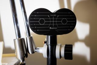
Trademarked and manufactured by Biomagnetic Technologies, a Sorrento Valley firm that is the only commercial supplier of multichannel MEG instruments in the world, the machine sits inside a specially shielded room that screens out the magnetic noise of day-to-day living.
Fourteen sensors called SQUIDs sit inside supercooled helium to detect the faint magnetic signals. Data are processed by computer into schematic brain maps, and multiple readings allow researchers to triangulate to determine the source of specific signals.
Magnetoencephalography promises to build on the knowledge of the brain gained through EEGs since they were first done in 1929.
For instance, researchers at New York University used MEGs to show that the auditory center, located at the bottom of a brain fissure above the ear canal, uses different groups of neurons to “hear” different pitched tones. Higher-frequency tones lie deeper within the fissure than do lower frequencies, said Samuel J. Williamson, a professor of physiology and biophysics at NYU Medical Center.
“It’s a very nice map that’s laid out very much like the surface of a piano. The distance from low C to middle C is the same as the distance from middle C to high C,” Williamson said.
Furthermore, more recent magnetic readings have hinted at how the brain pays attention to some sounds while ignoring others, he said. When a pair of songs are played simultaneously, the auditory neurons respond to both, but not with the same intensity.
“We find that when you pay attention to one of these tunes, the level of nerve activity is substantially enhanced compared to the response to the other tune,” Williamson said. “Yet it’s clear that we are processing both streams of information nevertheless.”
It is MEG’s ability to relate the brain’s well-known structure to its functioning that is the technique’s greatest potential as a research tool, Scripps’ Polich said.
“For the first time, you have the technological tool to determine what part of the brain does what when. With the MEG we’ll be able to say, ‘That’s happening now in this particular locus,’ ” he said. “You can get the ability to actually begin to specify precisely where thought occurs. That is just going to revolutionize psychology and psychiatry and anything that looks at mental operations.”
Relating structure to function also will be useful to medicine. Techniques such as CT scanning with X-rays and magnetic resonance imaging, which bombards the body with magnetism rather than measuring what it emits, both result in detailed structural maps of what a patient’s brain looks like. MEGs would help construct a three-dimensional map of how it functions.
At NYU Medical Center, researchers are beginning a project to determine how Alzheimer’s disease alters brain function, as revealed by MEG, Williamson said.
An answer might provide an easy way to diagnose Alzheimer’s, for which no routine test exists. It can only be positively confirmed by brain biopsy or, more commonly, by autopsy.
“Ten years from now, if it keeps going like the progression has been in the past, you’ll be able to bring in your grandmother and I think I’ll be able to tell you, ‘This is Alzheimer’s,’ as opposed to something else,” Polich said.
At the Veterans Administration Medical Center in Albuquerque, N.M., a Neuromagnetometer is being installed to study the brain function of partially paralyzed stroke patients.
“We know that before any kind of movement in a normal individual we see a change in electrical activity. What we’re trying to do is see if we can see those same fields in normal individuals magnetically,” said Kathy Haaland, a neuropsychologist there.
The change in electrical activity has two components, one that prepares the body to move and one that actually initiates the movement, she said. For stroke patients, it would be helpful to know which one is not functioning.
“If you could differentiate where the problem is, then there would be a better chance to develop rehabilitation strategies that related to one or the other,” Haaland said.
The Scripps research group, led by Dr. Floyd Bloom, head of preclinical neuroscience, later this year hopes to begin studies of the neuromagnetic patterns of Alzheimer’s patients, stroke victims and narcoleptics, Gallen said.
But the MEGs process is not without its problems, most of which have to do with the developing state of the technology.
The Scripps 14-channel magnetic detector is one of only two in clinical use worldwide (the other is at NYU), said Richard J. Faleschini, vice president for marketing and sales at Biomagnetic Technologies. The Biomagnetic machine at UCLA has seven sensors.
Even as what Gallen calls the “Corvette” of magnetoencephalography, the Scripps machine is laborious to use. Brain data that would take 10 minutes to gather on an EEG take about three hours with magnetoencephalography, and as yet there is no easy way to keep patients comfortable throughout the long process.
In addition, the machinery can be aimed only at certain angles, and software is still being refined to take account of different patient’s head shapes. Researchers have to figure out how to deliver visual and other stimuli to subjects without introducing magnetic “noise” into the specially shielded room where the machine is located.
The devices so far are so expensive--$900,000 for the Scripps machine--and have such narrow uses that some of the researchers doubt that they will ever be widely available in U.S. hospitals.
Perhaps the most serious long-term limitation for research comes because the brain’s magnetic fields are too weak for the technique to plumb the deepest parts of the brain.
Still, scientists are optimistic that the bugs will be worked out. They predict MEGs will be generally useful as a diagnostic tool within five years.


